|
Pulmonary Hemosiderosis
The Common Vein Copyright 2008
Definition
pulmonary hemosiderosis
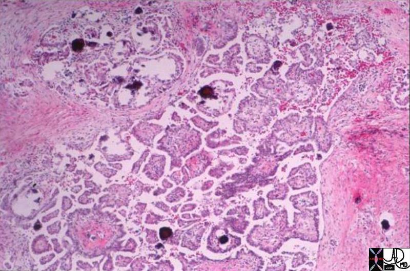
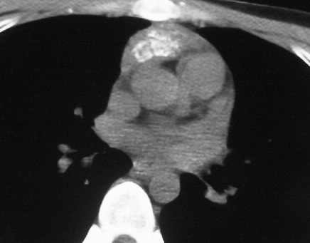
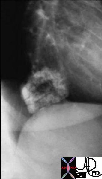
probale Pulmonary Hemosiderosis and Calcified Nodules Secondary to Mitral Stenosis
|
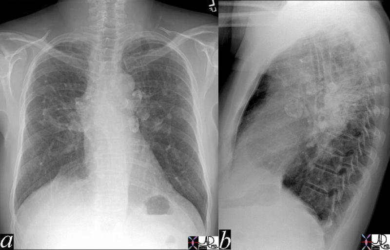
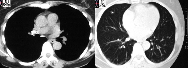
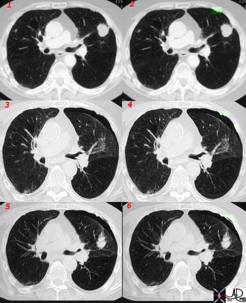
| 76286c05 elderly lady with dyspnea heart cardiac left atrium LA calcified LA s/p Carpentier rings in mitral valve tricuspid valve and AVR aortic valve replacement rheumatic heart disease RHD mitral stenosis fx interstitial lung disease ILD reticulonodular pattern dx probable pulmonary hemosiderosis with calcified or ossified nodules scout CXR CTscan Courtesy Ashley Davidoff MD |
probale Pulmonary Hemosiderosis and Calcified Nodules Secondary to Mitral Stenosis
|
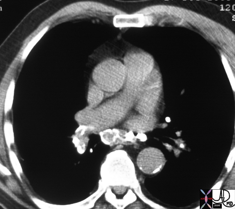
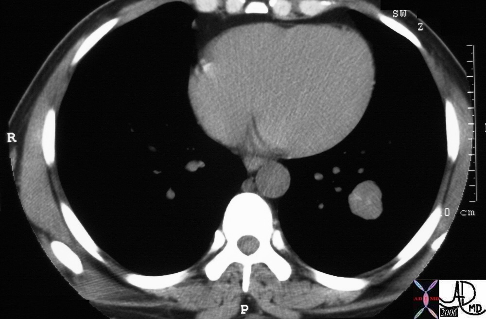
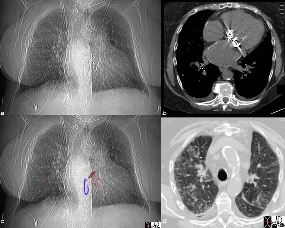
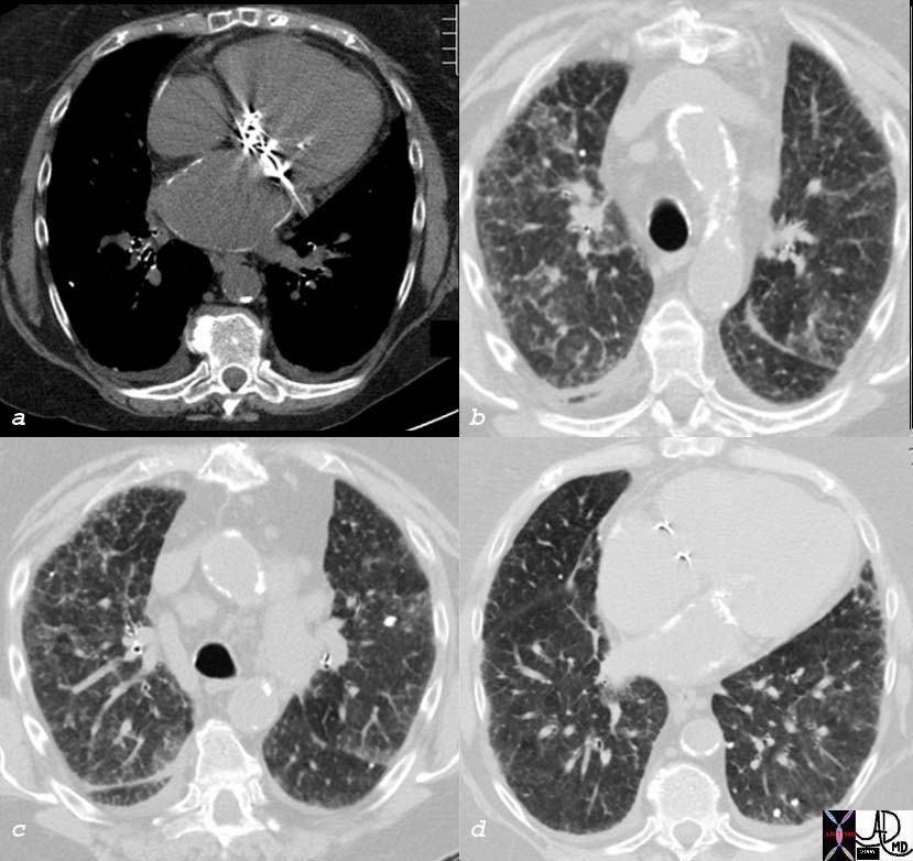
| 76286c07 elderly lady with dyspnea heart cardiac left atrium LA calcified LA s/p Carpentier rings in mitral valve tricuspid valve and AVR aortic valve replacement rheumatic heart disease RHD mitral stenosis fx interstitial lung disease ILD reticulonodular pattern dx probable pulmonary hemosiderosis with calcified or ossified nodules scout CXR CTscan Courtesy Ashley Davidoff MD |
References
eMedicine Singh V Mitral Stenosis
eMedicine Napchan G Pulmonary hemosiderosis
|