Copyright 2007
Introduction
Heart
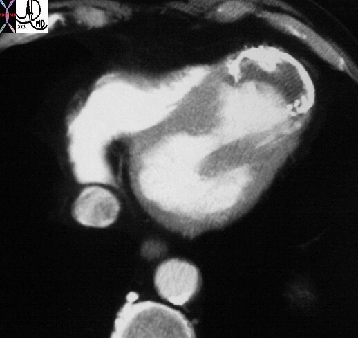
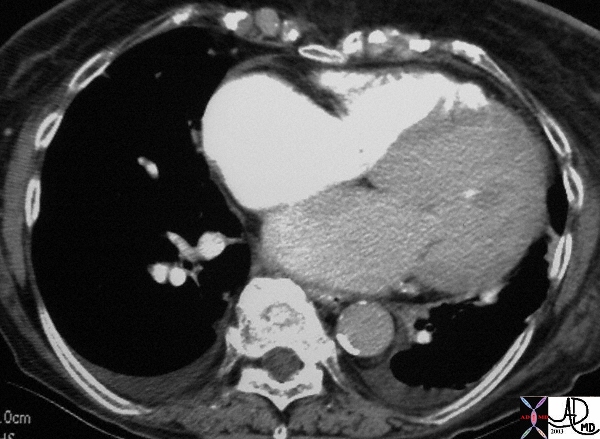
| 15401 |
| This CTscan with contrast through the heart shows heavy calcification of the LAD as well as the circumflex coronary artery as well as calcification of the aortic annulus. Courtesy Ashley Davidoff MD. 15401 code cardiac heart artery coronary calcification calcium calcified CAD atherosclerosis aortic sclerosis annulus imaging radiology CTscan AO aorta |

| 15527 |
| 15527 heart pericardium fx calcification serous pericardium fibrous pericardium dx calcific pericarditis probable viral in origin imaging radiology CTscan Courtesy Ashley Davidoff MD |
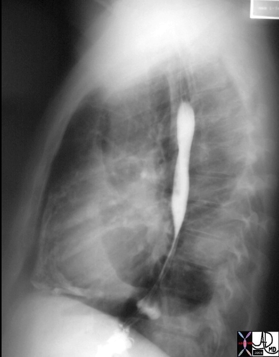
| 29157 |
| 29157 hx 55M with cough fx heart pericardium epicardium visceral pericardium parietal pericardium fx calcification dx calcific pericarditis probable viral virus imaging radiology plain film CXR Courtesy Ashley Davidoff MD |
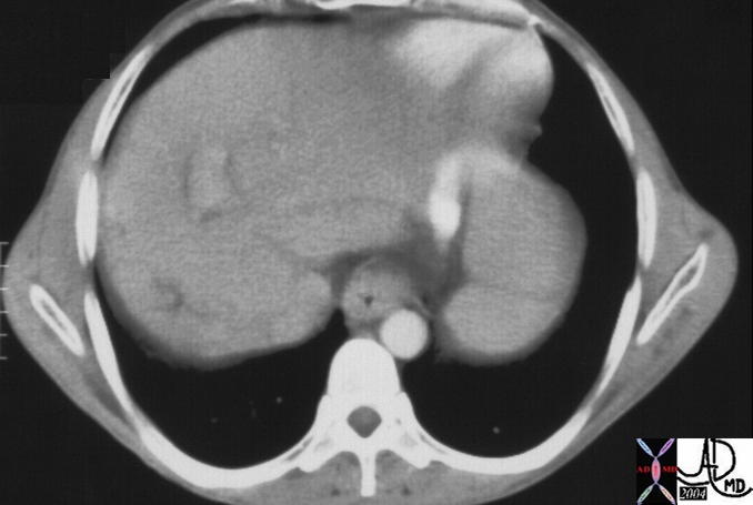
| 15528 |
| 15528 heart + pericardium + fx calcification + amyloid + imaging radiology CTscan infiltrative amyloidosis |
heart + pericardium + fx calcification + amyloid + imaging radiology CTscan infiltrative amyloidosis
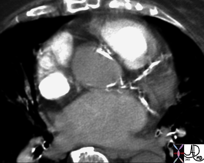
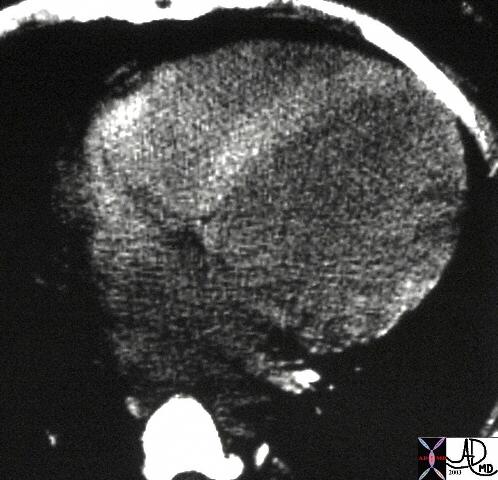
| 30472 |
| This cross sectional CT image of the heart shows calcified apex of the left ventricle associated with thrombus, characteristic of an LV aneurysm. The cause is almost certainly secondary to coronary artery disease and ischemic heart disease with secondary myocardial infarction. Courtesy Ashley Davidoff MD. 30472 code heart LV apex IHD CAD aneurysm calcification calcified MI cardiac imaging radiology CTscan |
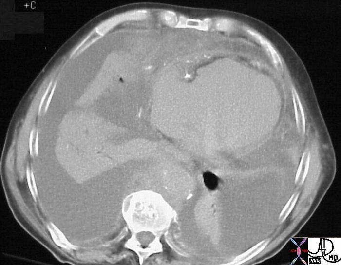
| 29600 |
| This single cross sectional view of the heart through the LV shows apical myocardial dystrophic calcification. The pericardium can be seen as a fine soft tissue density between the two layers of fat – the epicardial fat (inner layer) and the pericardial fat. This finding is diagnostic of a previous MI and may represent dystrophic calcification in the myocardium or within thrombus in an aneurysm of the apex. Courtesy Ashley Davidoff MD. 29600 code CVS cardiac heart MI apex calcification calcified IHD myocardium cardiac imaging radiology CTscan |

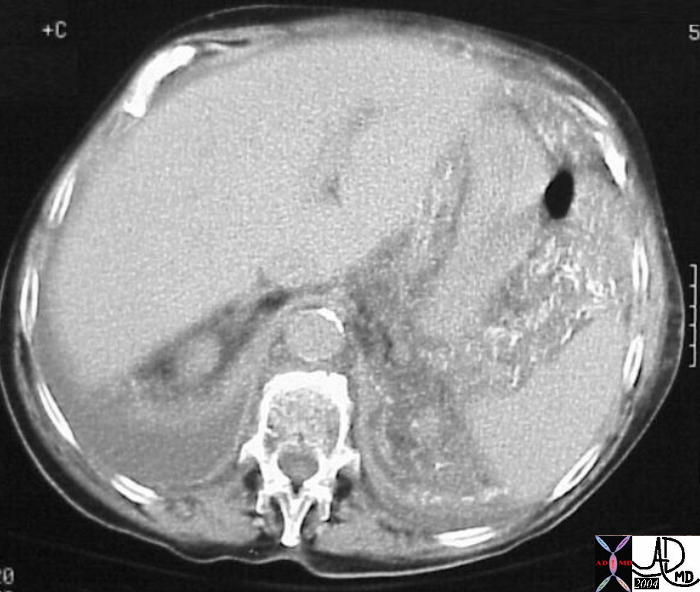
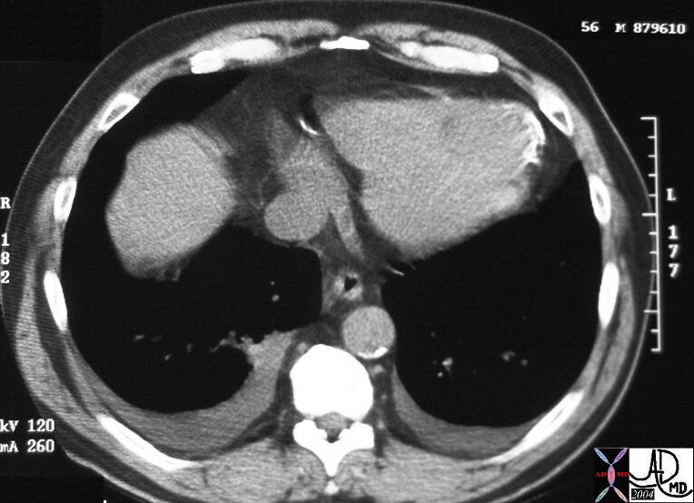
| 29601 |
| This single axial CT image shows a faint lucurvilinear lucency in the apical myocardium. This lucency is thought to be a fatty deposition in infarcted myocardial tissue. The calcification in the annulus is premature and unusual for this 56 year old male patient. Note the small bilateral pleural effusions. Courtesy Ashley Davidoff MD. 29601 code CVS cardiac heart MAC apical fat MI cardiac imaging radiology CTscan |
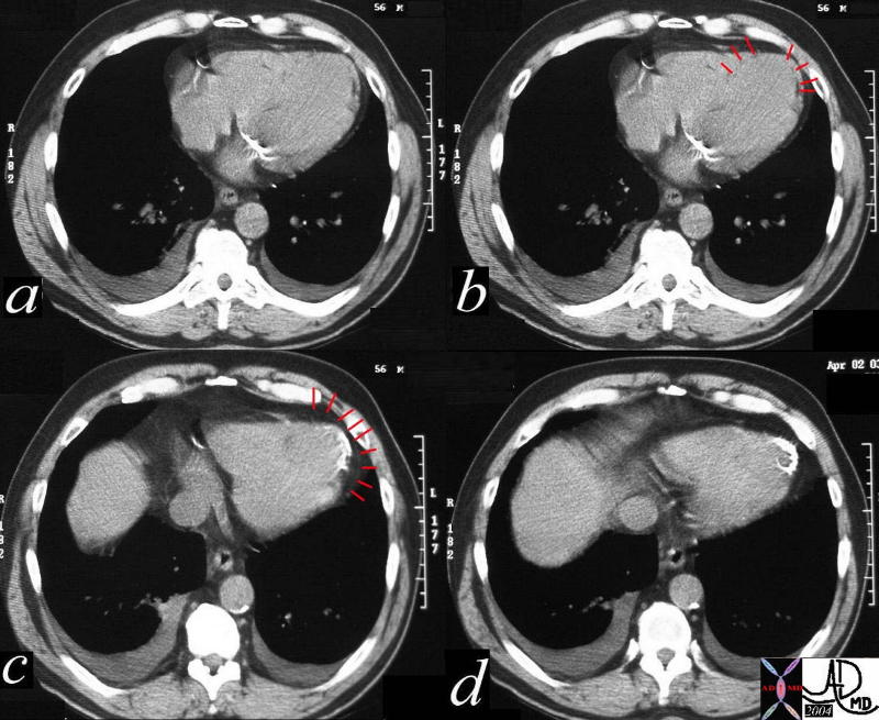
| 29601c01 |
| This series of CT image shows a faint curvilinear lucency in the apical myocardium (a,b) as well as myocardial calcification. The lucency identified by red lines in b, is thought to be a fatty deposition in infarcted myocardial tissue, and the calcification represents dystrophic calcification in the necrotic tissue. Note that the calcification is not in the pericardium which is identified by the red markers in c. The pericardium is suurounded by pericardial at on the outside and epicardial fat on the inside. Included in the differential diagnosis is an apical aneurysm with calcification in clot. The calcification in the annulus is premature and unusual for this 56 year old male patient. Note the small bilateral pleural effusions. Courtesy Ashley Davidoff MD. 29601c01 code CVS cardiac heart MAC apical fat MI calcium myocardium apex cardiac imaging radiology CTscan radiologists and detectives |
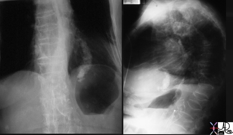
32744c01 |
| 32744c01 heart mitral valve mitral annulus mitral annular calcifcation MAC aortic caicifications osteopenia bone thoracic spine kyphosis compression fracture CXR plain film of the chest IVC filter Davidoff MD |
| 26320 |
| The chest CT is taken through the heart with the contrast phase in the right sided structures. A focal calcification in the cavity of the left ventricle (LV) is seemingly related to a focal hypodensity of the paillary muscle and may represent a calcific nodule on the chotdae or papillary muscle tip. Courtesy Ashley Davidoff MD 26320 code LV papillary muscle calcification chordae tendinae cardiac imaging radiology CTscan |
| 24672 |
| This is a CT of the chest at the level of the heart filmed on narrow windows showing a dense ventricular septum. The autopsy specimen showed calcific myocarditis. Courtesy Ashley Davidoff MD 24672 code heart septum dense calcific calcified calcification myocarditis inflammation cardiac imaging radiology CTscan |
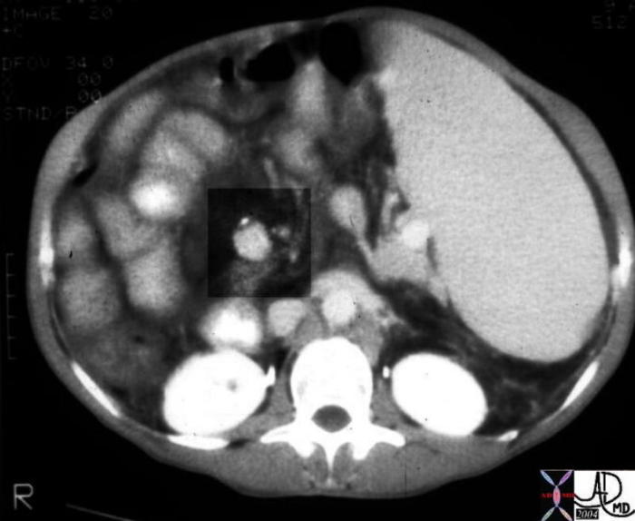
| 15529 |
| 15529 abdomen + gastrolienal ligament +spleen stomach fx calcification + amyloid + imaging radiology CTscan amyloidosis infiltrative |
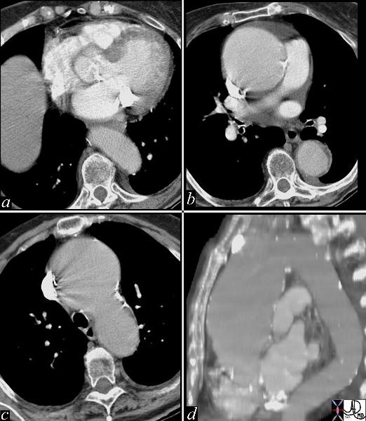
| 19426c |
| This series of images are from a CTscan showing an ascending aortic thoracic aneurysm. There is evidence of heavy calcification of the aortic valve (aortic sclerosis), an aneurysm confined to the ascending aorta (b,c,d), and tortuosity of the descending aorta (d). The cause for the aneurysm is probably a combination of systemic hypertension, aortic stenosis and atherosclerotic degeneration of the wall. Courtesy Ashley Davidoff MD 19426c code CVS thorax aorta ascending aneurysm MAC aortic sclerosis CTscan |
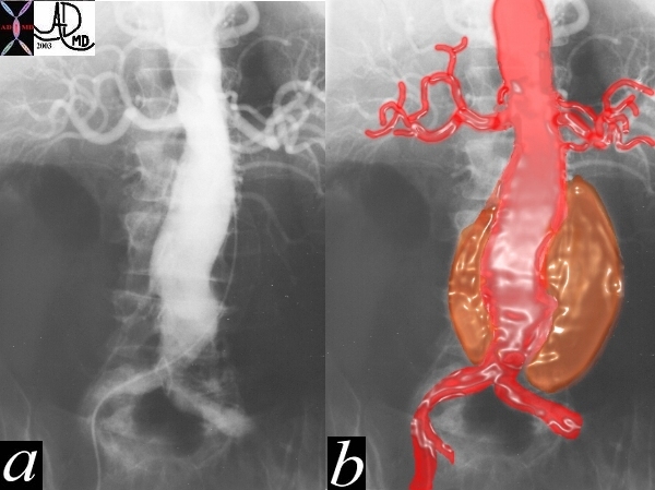
| 22734 cW02 |
| This angiogram of the abdominal aorta shows a widened infrarenal aorta. At first glance the lumen of the aorta appears normal, but a faint curvilinar calcification of the true wall can be seen to the patients left in the first image. The second image (b) reveals the true size of the aneurysm. Courtesy Ashley Davidoff MD 22734 cW02 codeCVS aorta artery abdomen aneurysm AAA |
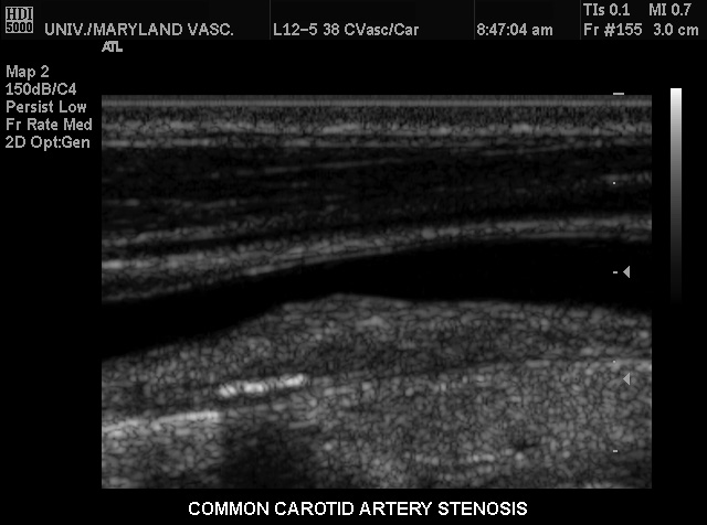
| 33289 |
| This conventional gray scale US of the neck shows a common carotid artery heaped up plaque on the far wall causing stenosis of moderate degree. Note also the linear area of strong echogenicity on the far wall associated with shadowing consistent with a calcification in the wall. These findings are characteristic of atherosclerotic plaque. Courtesy Philips Medical Systems 33289 |
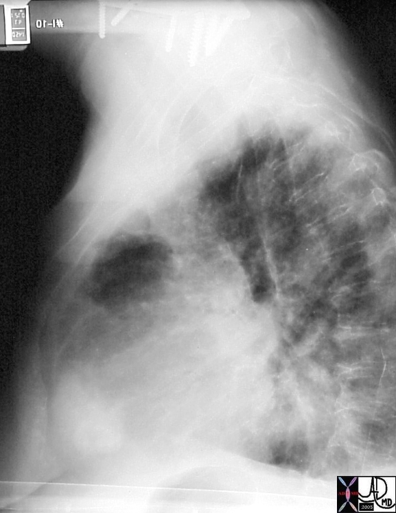
| 15537b01 |
| 15537b01 hx 75F with left shoulder pain heart fx calcification ddx question calcific pericarditis probable dx metastatic chondrosarcoma imaging radiology plain film Courtesy Ashley Davidoff MD |
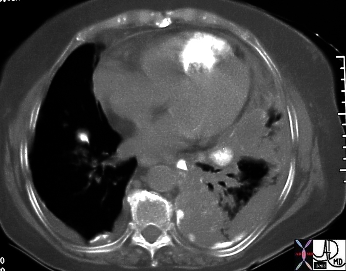
| 15537b03 |
| 15537b03 hx 75F with left shoulder pain heart lung fx calcification fx parenchymal infiltrate fx pleural effusion dx small pulmonary infarct small Hamptons hump dx metastatic chondrosarcoma metastasis osteogenic sarcomatous malignant pulmonary emboli embolus imaging radiology CTscan Courtesy Ashley Davidoff MD |
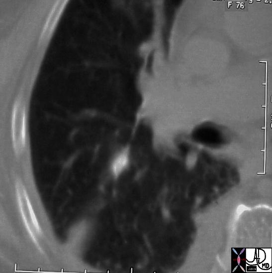
| 15537b04d |
| 15537b04d hx 75F with left shoulder pain heart lung fx calcification fx parenchymal infiltrate fx pleural effusion dx small pulmonary infarct small Hamptons hump dx metastatic chondrosarcoma metastasis osteogenic sarcomatous malignant pulmonary emboli embolus imaging radiology CTscan Courtesy Ashley Davidoff MD |
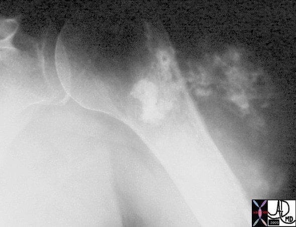
| 15537b07 |
| 15537b07 hx 75F with left shoulder pain heart left humerus left shoulder fx cartilagenous popcorn type calcification dx chondrosarcoma chondrogenic sarcoma of the shoulder with metastasis to the RV right ventricle malignant imaging radiology plain film Courtesy Ashley Davidoff MD |
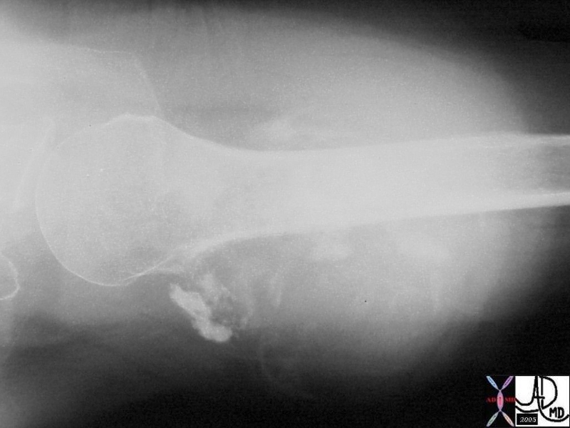
| 15537b08 |
| 15537b08 hx 75F with left shoulder pain heart left humerus left shoulder fx cartilagenous popcorn type calcification dx chondrosarcoma chondrogenic sarcoma of the shoulder with metastasis to the RV right ventricle malignant imaging radiology plain film Courtesy Ashley Davidoff MD |
Veins

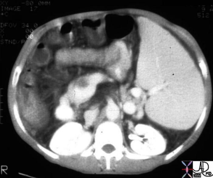
Massive Splenomengaly and….. |
| 00153 spleen enlarged splenomegaly portal hypertension vein SMV superior mesenteric vein fx calcification dx schistosomiasis schistosoma mansonii hepatic fibrosis cirrhosis imaging radiology CTscan |
| Massive Splenomengaly and calcification in the Portal Vein |
| 00153 spleen enlarged splenomegaly portal hypertension vein SMV superior mesenteric vein fx calcification dx schistosomiasis schistosoma mansonii hepatic fibrosis cirrhosis imaging radiology CTscan |
Liver – Bands of Fibrosis |
| 00153a01 liver + deformity + fx linear hypodense bands of fibrosis dx schistosomiasis + schistosoma mansonii imaging radiology CTscan infection cirrhosis portal hypertension |
More Thrombus |
| 00154 spleen enlarged splenomegaly portal hypertension vein SMV superior mesenteric vein thrombosis fx dx schistosomiasis schistosoma mansonii hepatic fibrosis cirrhosis imaging radiology CTscan |
|
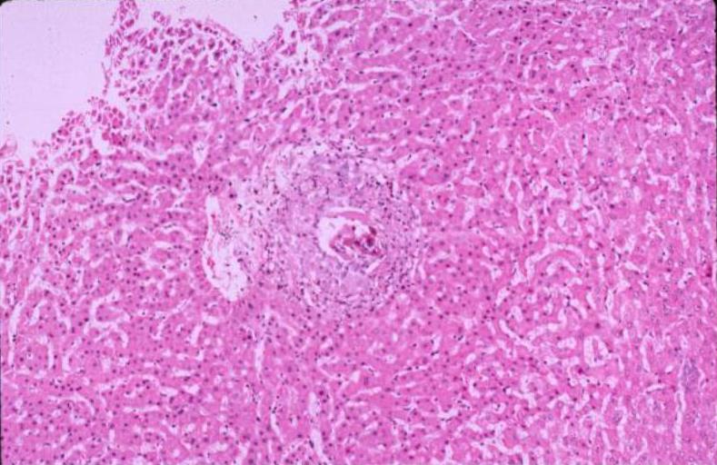
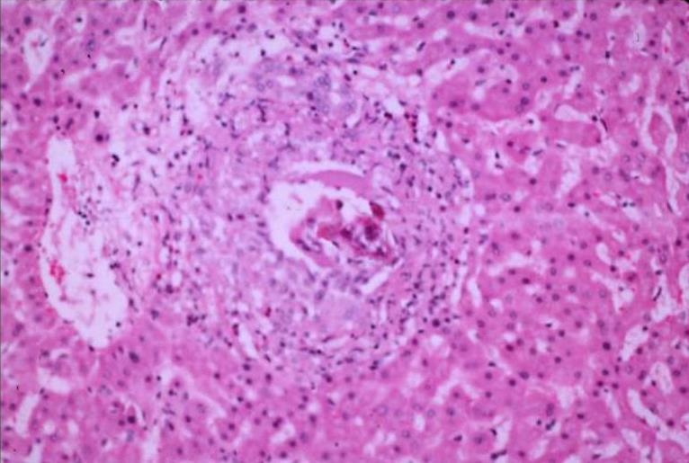
Granuloma in the Portal Vein with the Egg in the Center |
| 02273 02275 hx young man from Puerto Rico liver schistosomiasis schistosoma hematobium egg histopathology Courtesy Barbara Banner MD |
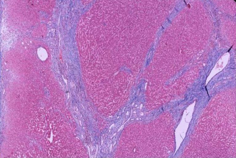 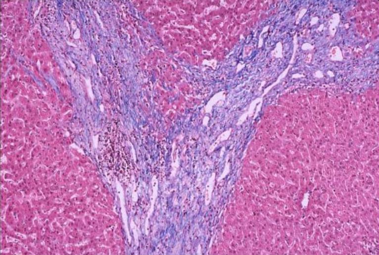
Bands of Fibrosis |
| 02270 02272 hx young man from Puerto Rico liver schistosomiasis hepatic cirrhosis histopathology Courtesy Barbara Banner MD
portal fibrosis stand out , termed as “pipestem” fibrosis.
|
