1827 –
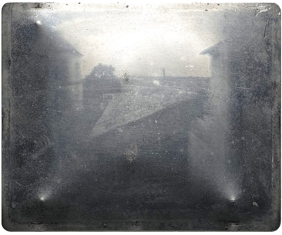
The earliest photograph available of a building and a tree taken by Frenchman Joseph Niépce later to become Nicephore Niepce was in 1827 and it took 8 hours for exposure and who knows how long to develop.
Courtesy Wiki
8 hour exposure
1895
In 1895 Wilhelm Roentgen discovered the use of X-Rays
Courtesy Wikipedia
15minutes
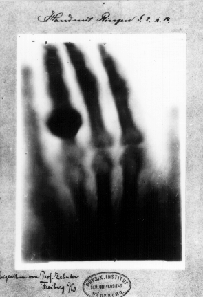
Courtesy Wikipedia
1975 Nobel prize
Award Winner –
Although computerized tomography (CT) – or computerized axial tomography (CAT) – scan is associated with science and technology, many would be surprised to learn that its humble beginnings were through a company – EMI that was successful in promoting the Beatles

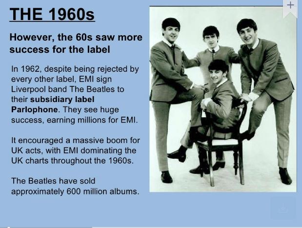
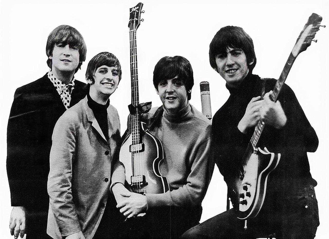
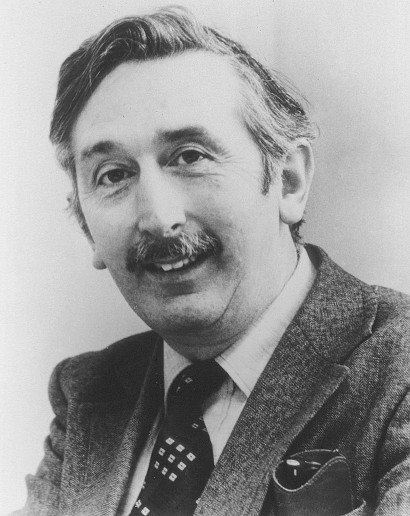
Their record company was EMI for whom Godfery Hounsfield worked and it was through their success as a record company that they were able to finance the research of Godfrey Hounsfield
could possibly be rooted in rock and roll. Specifically, the creation of this incredible technology has been widely attributed to the success of The Beatles in the 60s.
1972 CT scan
Wiki
South African American physicist who won the 1979 Nobel Prize in Physiology or Medicine (along with Godfrey Hounsfield) for his work on X-ray computed tomography (CT)
Wiki
Allan Cormack, a physicist, developed a
mathematical process for calculating the distribution of x-ray attenuation values throughout a simulated body section.
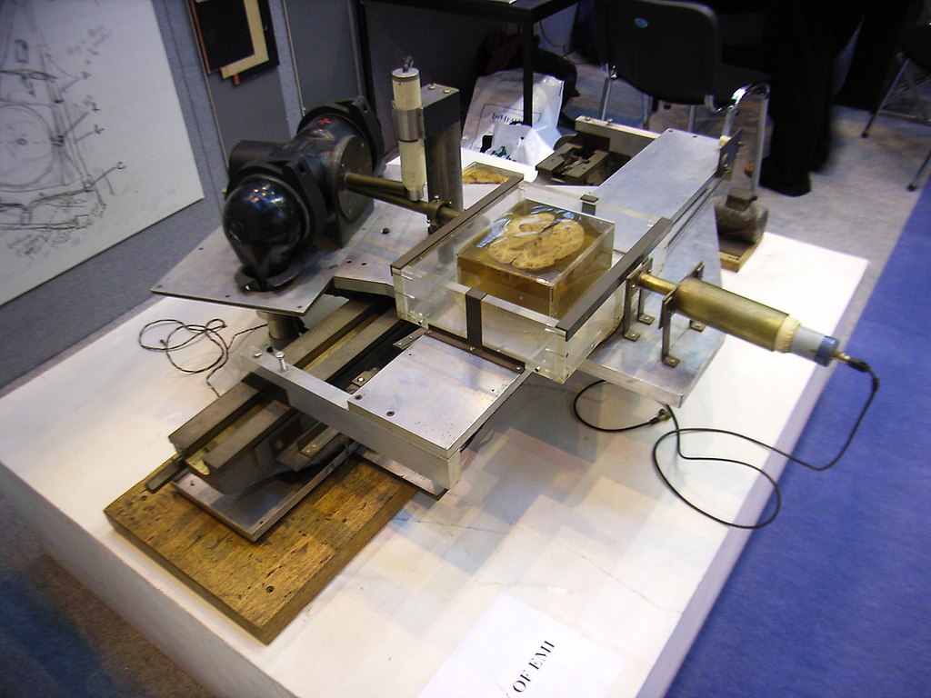
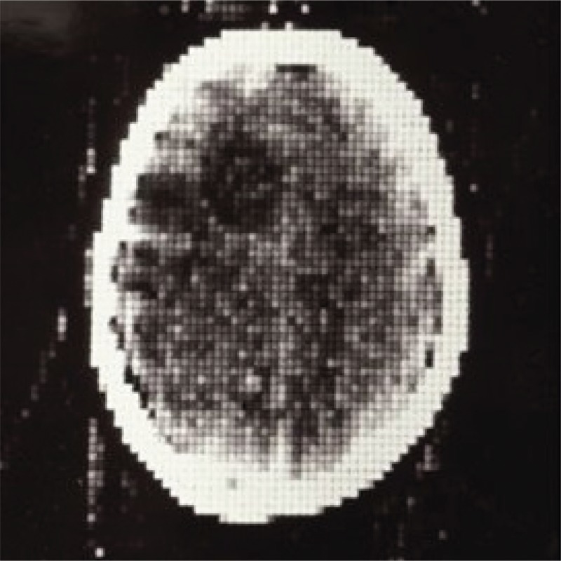
The first clinical image was taken by Hounsfield in 1972 on an EMI scanner when he scanned the brain of a cow – It took several hours to scan a single slice and several days to produce the image. The graininess and lack of clarity in both images is similar with poor resolution , due to the long exposure times.
-
1972 First Clinical Scan
- 30 minutes for the scan,
- a drive across town with the magnetic tapes to EMI,
- 2.5 hours processing the data on an EMI mainframe computer
- capturing the image with a Polaroid camera
- racing back to the hospital
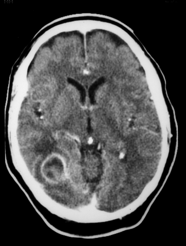
- Result
-
- a cystic mass about the size of a plum in the right frontal region
-
Progress in both the fields of photography and CT scanning have evolved principally due to the speed with which the image is acquired. The current advances depend heavily on both the speed as well as the digital method. Thus advance, particularly in the world of biology, where things are on the move, have paralleled the progressive sophistication of computers.
1973 Robert Ledley – a whole-body scanner
Single Row of Detectors 5,7, 10mm
2 Seconds with 6 second interscan delay
The gantry or the doughnut through which the patient is scanned – originally contained a singe row of detectors and the slice thickness thus depended on the collimation of the beam and the size of the detectors in general 5,7, 10mm
The gantry was able to rotate one way (say clockwise) and then had to reverse direction and rotate counterclockwise to uncoil the cables that were involved in delivering the high voltage power, and the data. Each 2-second scan was separated by a 6-second interscan delay (needed to reorient the x-ray source-detector assembly within the gantry to prevent entanglement of the cables) The patient also needed to be moved to acquire the next slice. If the patient happened to move, breath or a heart beat occur between the move, then the slices did not line up. The brain was the most photogenic of them all – (mirror mirror on the wall ), because it was able to stay still for the camera – but peristalsis, respiratory motion and heart beats made moving targets in the rest of the body.
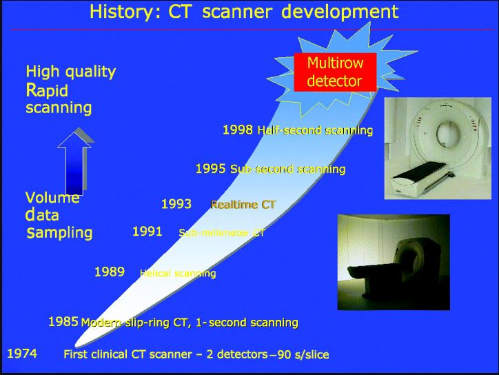
Slip Ring Technology late 1970’s
The application of slip ring technology, which enabled a continuous motion of the gantry in helical fashion allowed the advances necessary First slip-ring CT system was introduced by Varian in the 1970s,
1980’s Spiral Scanning 1988
Sieman’s capitalized in 1988 by introducing helical or spiral scanning technology.
1992 Dual Row of Detectors
A second breakthrough was introduced by Elscint who introduced a second row of detectors in 1992, which became 4 in 1998, and now we are up to 256. Thus we have to in the world of analog photography speed and resolution are major factors With the advent of digital techniques speed and resolution – now measured in amount of data strength speed and power of the computer
The first computed tomography machine that was commercially available in the United States (about 1972) used an 80 by 80 pixel matrix. A cross-sectional image of the brain
Ashley Davidoff thecommonvein.net 14917
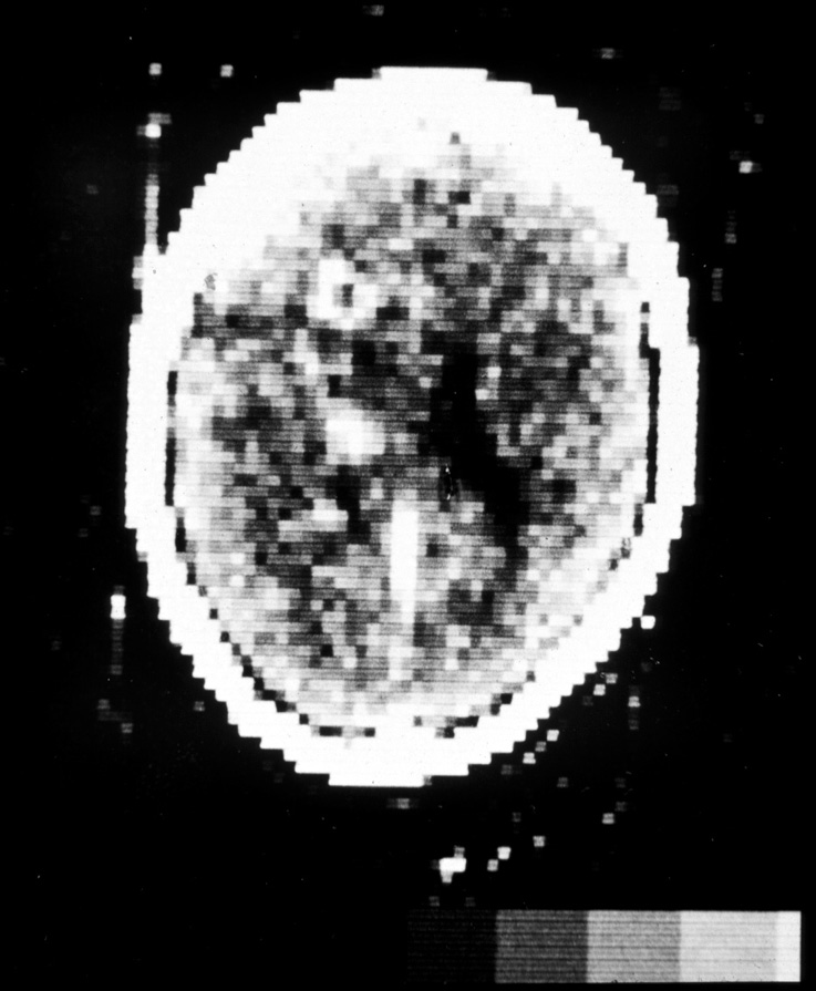
A similar cross section of the brain done on a more modern 512 by 512 matrix machine (14917 )
‘
Godfrey Newbold Hounsfield, a British scientist, and Allan M. Cormack, a South African physicist, won the Nobel Prize in physiology or medicine in 1979 for their development of computer assisted tomography.
1963
Cormack AM
1972
Hounsfield GN
EMI Mark I
first commercial scanner required 41/2 minutes to complete a
brain scan
first-generation
line integral collected using an
x-ray tube and a
single detector
second-generation scanners
line integral collected using an
x-ray tube and a
multiple detectors
third-generation systems
fan beam x-ray source
large array of detectors
fourth-generation systems
fan beam x-ray source
fixed detector array completely surrounds the patient”
Links and References
- Higgins E 50 years ago, the first CT scan The Conversation 2021
- Baker, H. 1993. “Historical Vignette: Introduction of Computed Tomography in North America.” American
Journal of Neuroradiology; Vol. 14, pp. 283-287. - See Mayo Clinic Newsletter Re Dr Baker a Radiologist
- TCV