Disease
The Common Vein copyright 2007
Introduction
We learned in an earlier part of the module that there is a universal equation that defines the interactions between objects.
Things interact with other things in a given environment at a moment in time resulting in a change. This change may be either good or bad – positive or negative.
When the change is bad – disorder results and the biological equivalent of disorder is disease.
There are about 12 basic disorders of disease.
1 Inflammation
2 Infection
3 Neoplasm amd other growth abnormalities
4 Mechanical disorders
5 Trauma
6 Metabolic disorders
7 Circulatory
8 Immune disorders
9 Iatrogenic
10 Inheritd
11 Idiopathic
12 Functional
13 Healling that has as a consequence a degree of disorder
Inflammation
Inflammation is th body’s response to an injurious or noxious agent. This noxious agent by virtue of its perceived malintent generates a protective response by the body. This response initially consists of two a vascular response and a cellular response. The vascular response results in an increase in blood flow to the region resulting in both an increase in fluid and an increase in leukocytes to the interstitial space at the site of injury.
The increase fluid in the region dilutes the noxious agent and the infiltration of leukocystes serves as a direct atttempt at destroying the invasive agent.
The damage caused by both the invasive agent and to variable extent the inflammatory response requires repair which is also instituted by the inflammatory response. Initially this consists of fibrin deposition with subsequent evolution of a longer lasting fibrotic change.
Celsus 200 years ago – rubor calor dolor tumor laesio function (Virchow (1821 -1902
Hyperemia calor
 Radiation Hepatitis – Hyperemia Radiation Hepatitis – Hyperemia |
| The straight line and the hyperemia are characteristis features radiation induced change
22975 Courtesy Ashley Davidoff MD |
Infection
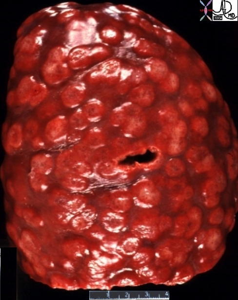
Splenic Granulomas |
| 21327a 21324 spleen + multiple splenic nodules dx non specific splenic granulomas + grosspathology inflammation infection |
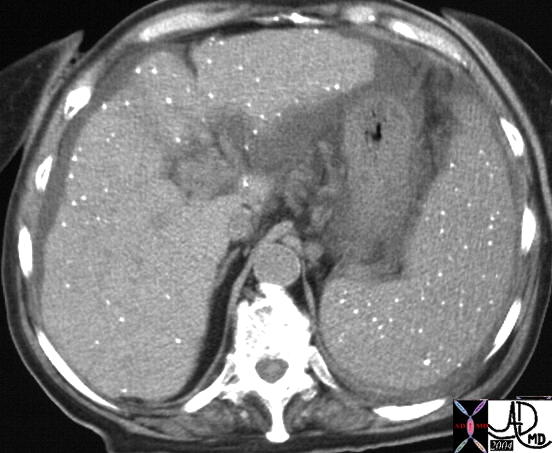
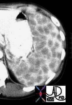
| Tuberculosis – Chronic Granulomatous Disease |
| 89 year old female with a history of bovine TB 20752 spleen + liver splenic nodules + fx calcification + calcified hepatic granulomas TB + imaging radiology CTscan C- infection granulomatous disease |
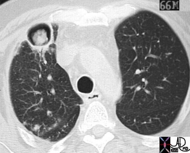
Aspergillus – A Fungal Ball |
| 20760 lung + fx mass + fx air + soft tissue aspergillus + imaging radiology CTscan aspergilloma fungus fungal infection pulmonary systemic fungemia with involvement of liver and spleen code air crescent sign |
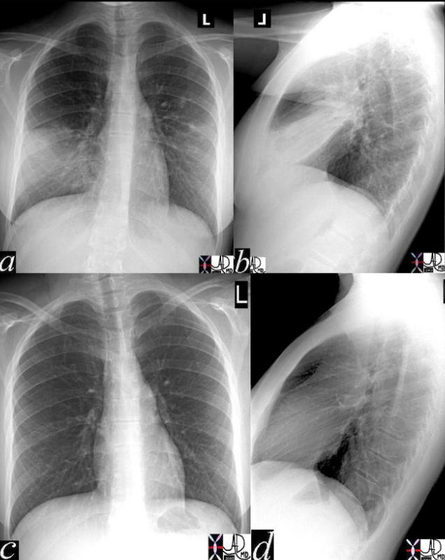
Before and After Antibiotic Therapy |
| 41819c02 Courtesy Ashley Davidoff MD medical students code chest CXR imaging lung plain film pneumonia radiology resolution |
Malignancy
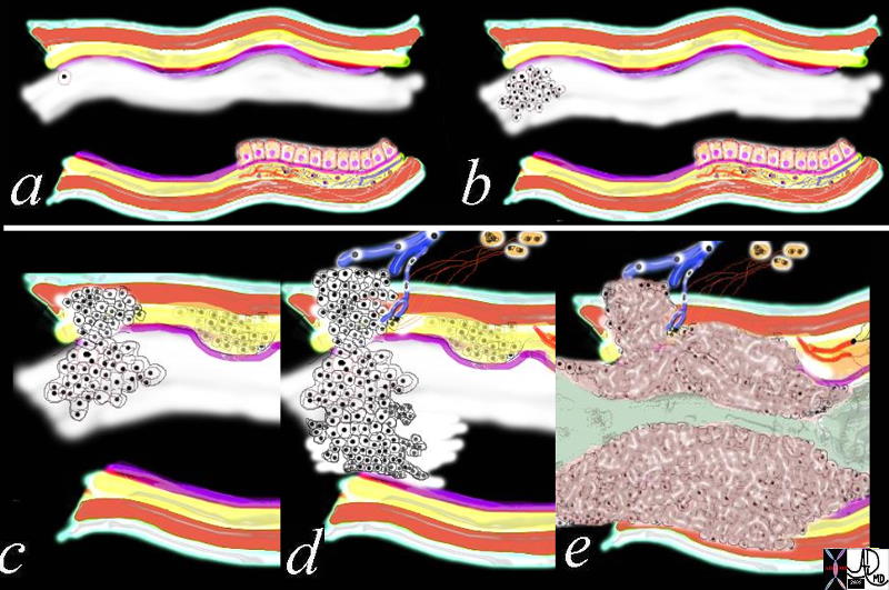
Growth of a Malignancy |
| 32354c01.800 tube colon trachea bronchus small bowel bile duct epithelium neoplasm benign malignant malignancy epithelial cell multiply multiplication growth mucosa submucosa muscularis serosa adventitia vein lymph node circumferential metastasixe metastasis metastases narrowing stenosis obstruction complication cancer uncontrolled growth Davidoff drawing Davidoff art Davidoff tube Davidoff MD |
Malignancy Occupying Space – The Faces of Malignancy
Spiculated Masses
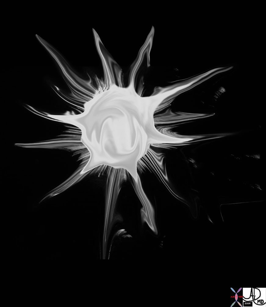   Stellate Stellate |
| 69595b05.800 Davidoff art star stellate corona radiata spiculated aggressive cancer carcinoma
70142.800 tree roots stellate shape Davidoff photography New Hampshire 46361b01 lung fx spiculated nodule dx carcinoma CTscan Courtesy Ashley Davidoff MD |
 Spiculated Lung Nodule Spiculated Lung Nodule |
| 46361 lung fx spiculated nodule dx carcinoma CTscan Courtesy Ashley Davidoff MD |
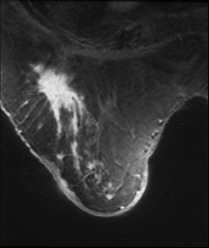 Spiculated Mass in the Right Breast Spiculated Mass in the Right Breast |
| This is an MRI of the right breast of a 78 year old patient with a remote history of invasive lobular carcinoma. The finding on the MRI is characterized by an enhancing spiculated mass. Recurrent carcinoma was present at pathology. Courtesy Priscilla Slanetz MD MPH 42977 |

 Infiltrative Malignancy
| Infiltrative Adenocarcinoma of the Lung |
| 46588 46581 lung fx consolidation dx adenocarcinoma of the lung malignancy cancer Davidoff MD |
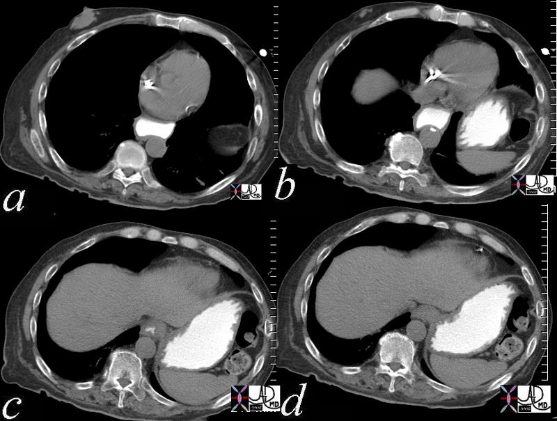
Malignancy – Vascular Invasion

| Vascular Invasion |
| 46579 liver hepatic vein fx tumor thrombus lung fx consolidation dx infiltrative adenocarcinoma of the lung with metastases to the liver and adrenal and invasion of the hepatic veins malignancy cancer Davidoff MD 46581 46588 46587 |
Malignancy – Vascular Encasement

Vascular Encasement |
| 46645c03.800 46645c04.800heart artery pulmonary trunk encasement right ventricle RV infundibulum dx carcinoma of the lung CTscan Davidoff MD |

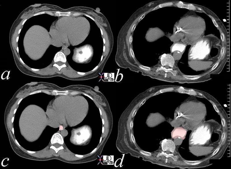
Malignancy – Space Occupying

| Squatters in the Left Lobe |
| 46587.800 liver bone vertebra fx mass space occupying disease dx liver metastases metastasis dx primary lung carcinoma malignancy cancer CTscan Davidoff MD |
Congenital Growth Disorders

| Pelvic Kidney |
| 46610c01 kidney pelvic kdney renal position ureterolthiasis ureterovesical junction stone UVJ stone ureteric stone fx hydronephrosis fx etravasation dx acute obstruction of the right ureter from a 2mm calcified ureteric stone in a patient with a pelvic kidney CTscan Davidoff MD |

Sinle Kidney Bicornuate Uterus |
| 46527c01 uterus kidney renal bicornuate uterus single kidney congenital abnormality congenital growth disorder USscan MRI Davidoff MD |
Mechanical Disorders – Obstruction
|
Acute Ureteric Obstruction |
| 46610c01 kidney pelvic kdney renal position ureterolthiasis ureterovesical junction stone UVJ stone ureteric stone fx hydronephrosis fx etravasation dx acute obstruction of the right ureter from a 2mm calcified ureteric stone in a patient with a pelvic kidney CTscan Davidoff MD |
Mechanical Disorder Fracture
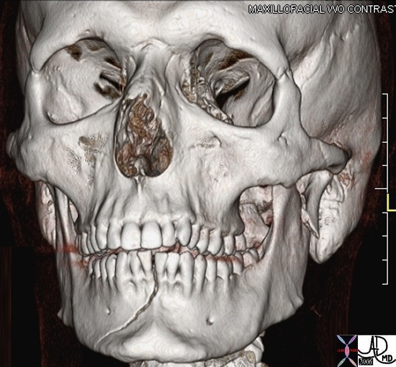
Fractured Mandible |
| 45669 bone maxillofacial mandible face cheek orbit frontal sinus teeth maxilla nasal bone concha zygomatic bone dx fracture of the mandible fracture of the left condyle TMJ skull CT scan Courtesy Ashley Davidoff MD 5star |
Mechanical Disorders – Degeneration

Wrath of Time |
| 46707c01 bone wrist carpals thumb degenerative changes osteoarthritis osteopenia age time normal vs abnormal X-ray plain film Davidoff MD |
Mechanical Disease – Loss of Elasticity
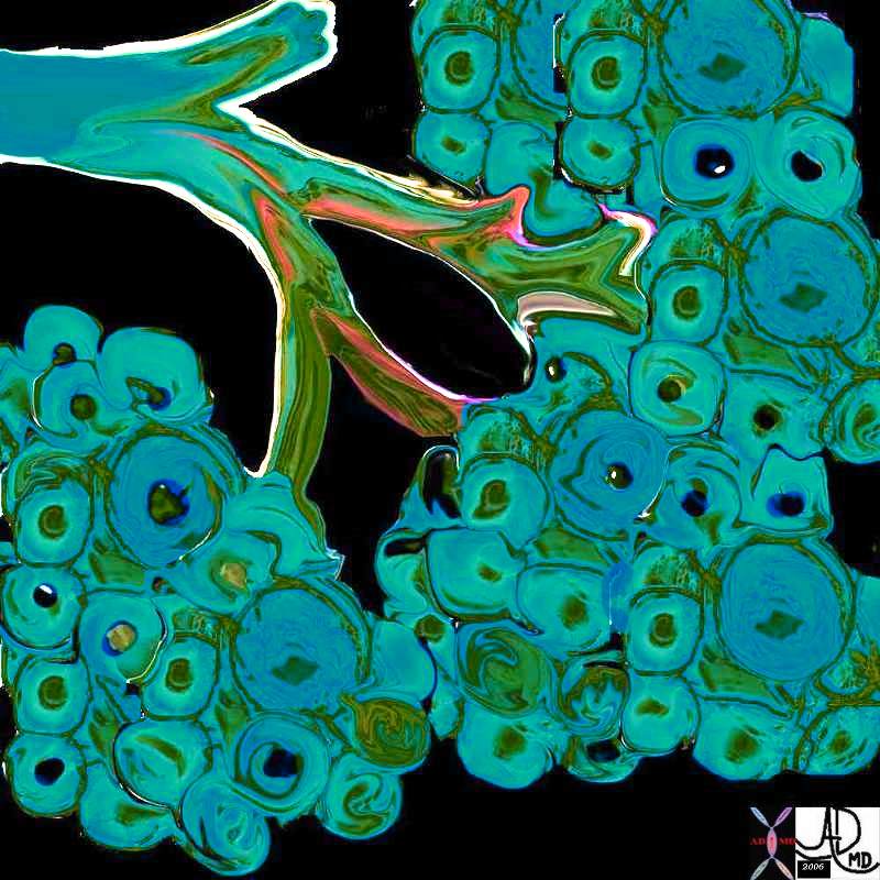 
Normal Alveoli of the Lung and Emphysema
|
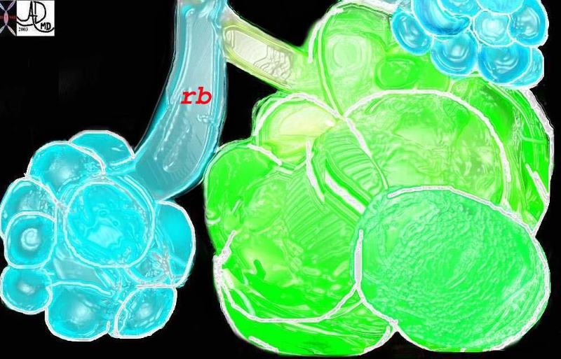
| This diagram illustrates the branching pattern of the tracheobronchial tree that extends from the bronchi to the terminal bronchioles transitioning into the alveoli via the alveolar sacs. Courtesy Ashley Davidoff MD 32645b04b04
This diagram shows alveoli and respiratory bronchioles that are too large due to loss of elasticity, so that air cannot be moved efficiently through them This is a diagram of emphysema causing hyperinflated lungs lung volumes 32645b01.800 Davidoff art |
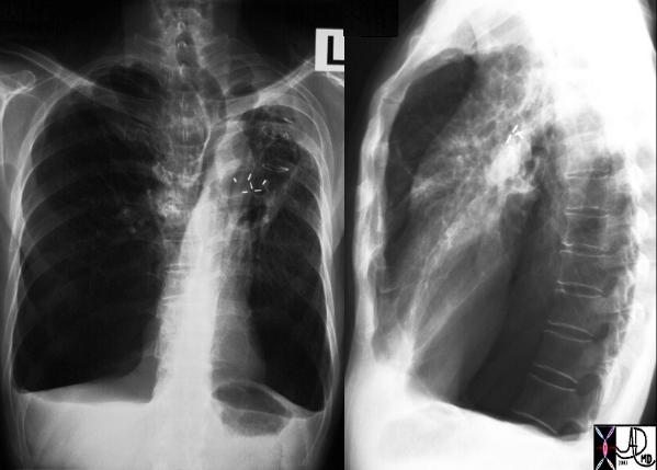
| Large lungs |
| A patient with hyperinflated lung volumes, COPD, and emphysema with surgical removal of a lung carcinoma from the LUL. Note how flattened the hemidiaphragms are and note the large retrosternal air space and the shapoe of the chest – like a barrel – called pectus carinatum – or pigeon chest. The lungs are literally so large that they are pushing the chest wall forward.
Courtesy Ashley Davidoff MD 30672c |
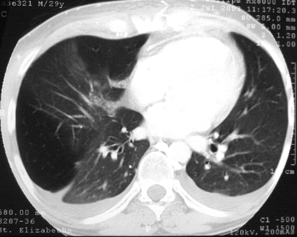
Segmental emphysema |
| This is a chest CT of a 29 year old man man who has segmental emphysema in the right lower lobe and midle lobe characterised by lucencies at 3 and 12 oclock where there has been air trapping in the alveoli. The left lung is normal This condition is called Swyer James syndrome Courtesy Ashley Davidoff MD. (30314 ) |
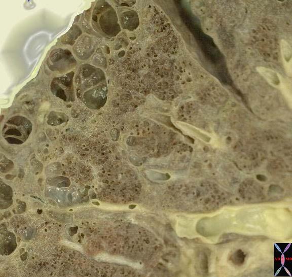
Emphysema |
| This is an image of an emphysematous lung. Note the larger air spaces where the septae between the alveoli, alveolar sac, salveolar ducts and respiratory bronchioles have been broken down. 19932e |
Circulatory Disorders

| Cardiogenic Shock Drowned Lung |
| 46813c01.800 chest lung pulmonary edema alveolar edema batwing distribution drowned lung dax cardiogenic shock normal CXR plain film of the chest Davidoff MD |

CHF – AVR |
| 46432c01 heart cardiac chest fx pulmonary congestion interstitial edema enlarged pulmonary arteries pulmonary arteriole to bronchiole ratio increased cardiomegaly fx enlarged Kerley B line thickened interlobular septa dx congestive heart failure CHF cardiac failure CTscan CXR plain film Davidoff MD 46425 46427 46428 46431 46432 46432c01 |

|
Skin CHF |
| 46786c02 skin heart cardiac breast fx left atrial enlargement LAE fx third spacing fx effusions fx skin edema cardiac failure CHF CTscan Davidoff MD |

Acute and Chronic Infarction |
| 49685C01 brain DWI occipital lobe fx vague hypodensity right occipital lobe with encephalomalacia and ex vacuo changes in the left occipital and posterior parietal region dx acute infarction right occipital lobe chronic infarction left occipItoparietal lobe a IR white matter disease vague increase in right b T2 gliosis left parietal c DWI bright acute right occipital d CT vague hypodensity right occipital old infarct left dx acute infarction right occipital lobe chronic infarction left parietal lobe MRI diffusion weighted imaging CTscan Courtesy Ashley Davidoff MD |

| Acute GI bleed |
| 46590 small bowel fx radioisotope leak into small bowel dx GI bleed hemorrhage Radioactibe labelled sulfur colloid Nuclear Medicine NMscan Davidoff MD |

| Acute Subdural Hematoma |
| 46633c01.800 brain cerebral meninges subdural hematoma blood SDH hemorrhage bleed acute blood fx acute hematoma s/p trauma CTscan Davidoff MD |

Hemorrhage – Ectopic Pregnancy |
| 46750c01.800 hx young female presentingwith vaginal bleeding for 2 weeks, hematocrit of 25, left lower quadrant pain, and LLQ mass HCG 7000 fx uterus endometrial stripe ovary fx mass echogenic cystic complex free fluid loculated fluid hemorrhage blood fx no intrauterine pregnancy dx ectopic pregnancy death Davidoff MD |

Hemorrhage |
| 46750c02.800 hx young female presentingwith vaginal bleeding for 2 weeks, hematocrit of 25, left lower quadrant pain, and LLQ mass HCG 7000 fx uterus endometrial stripe ovary fx mass echogenic cystic complex free fluid loculated fluid hemorrhage blood fx no intrauterine pregnancy dx ectopic pregnancy death USscan Davidoff MD |

Wedge Shaped Infarction |
| 46772c01.800 spleen splenic fx wedge shaped defects dx emboli dx systemic embolization dx splenic infarcts CTscan Davidoff MD |
Functional Disorders
Achalasia |
| 46561c01.800 esophagus stomach gastroesophageal junction fx enlarged esophagus megaesophagus fx dilated fx narrowing at the GE junction fx stenosis dx achalasia CTscan Davidoff MD |
| Normal and Abnormal |
| 46561c02.800 esophagus size normal dilated enlarged dx achalasia obstruction proximal dilation CTscan Davidoff MD |

| Aspiration |
| 46508c01.800 esophagus dysphagia fx large osteophyte causing difficulty with passing a scope fx aspiration barium swallow x-ray contrast Davidoff MD |
Healing

| Healing over 4 weeks |
| 46797 bone hand metacarpal fx fracture dx boxer’s fracture position alignment post pinning healinhg over 4 weeks osteoid X-ray plain film Davidoff MD Davidoff MD |
Death

| Life and Death based on size shape and Position |
| 46592c01 uterus OB pregnancy fetal demise spontaneous abortion shape size position heart rate USscan Davidoff MD death |

| Ectopic Pregnancy |
| 46767c01.800 hx young female presentingwith vaginal bleeding for 2 weeks, hematocrit of 25, left lower quadrant pain, and LLQ mass HCG 7000 fx uterus endometrial stripe ovary fx mass echogenic cystic complex free fluid loculated fluid hemorrhage blood fx no intrauterine pregnancy dx ectopic pregnancy death USscan Davidoff MD |