copyright 2007
Principles
BASIC X-RAY PHYSICS
A sound understanding of imaging techniques and the physical principles behind them enhances our ability to visualize and appreciate the internal structure and functions of the living body.
Diagnostic X-rays are generated by conversion of the energy acquired by electrons accelerated through an electrical field gradient in the kilovolt (kV) range
X-rays are very high frequency, with short electromagnetic waves
Diagnostically useful wavelengths are between 0.06nm and 0.006nm
An electrically heated filament (cathode) in an X-ray tube generates electrons that accelerate toward a tungsten target (anode) when a high voltage is applied to the tube
As electrons decelerate upon collision with the tungsten target, energy in the form of X-rays (which came at the expense of the kinetic energy of the electrons) is emitted
A beam of X-rays is created and can be aimed at a desired target such as a chest
About 1% of X-rays directed at the body reach the film to expose it
mA vs kVp
mA – milliamperes
Quantifies current from cathode to anode
Higher mA resulting in more electrons hitting anode per unit time leading to a larger quantity of x-rays
kVp – kilovolts peak potential
Quantifies voltage difference between cathode and anode
Higher kVp resulting in more “penetrating” x-rays
INTERACTIONS OF X-RAYS WITH MATTER
No interaction:
X-ray passes completely through tissue to imaging recording device
Complete absorption:
X-ray energy is completely absorbed by the tissue  no imaging information results
Partial absorption with scatter:
Scattering results in partial energy transfer to tissue energy and alters the trajectory of X-rays
FACTORS AFFECTING ATTENUATION
Tissue electron density
Dense material (bone, contrast dye) attenuate more X-rays than less dense material (muscle, fat, air)
Tissue thickness
Probability of scatter and interaction increases with tissue thickness (more attenuation)
X-ray energy (kVp)
Increasing kVp means greater beam penetration through tissue (less attenuation)
X-RAY ATTENUATION:BLACK WHITE AND SHADES OF GRAY
BONE
strongly attenuates X-rays (large attenuation coefficient), and presents radio-graphically in white
AIR
Weakly attenuates X-rays (small attenuation coefficient), and therefore presents radio-graphically in black
GRAY
Structural elements that attenuate the beam to a greater extent than air or are less attenuating than bone (i.e. soft tissues) show radiographically in various shades of gray

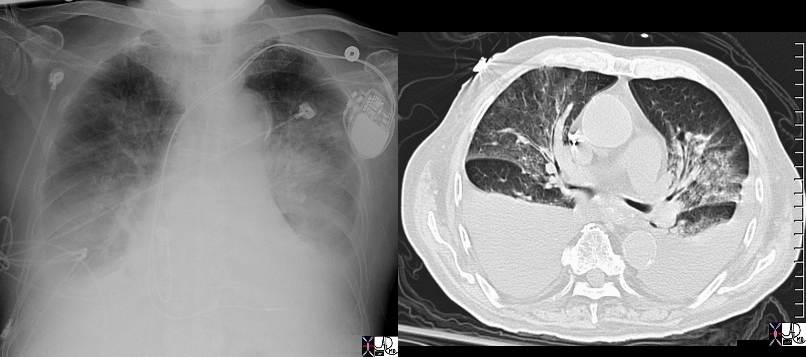
Batwing Shape of Perihilar Congestion in Acute CHF |
| 49451c01 heart cardiac batwing distribution afx interstitial and alveolar edema air bronchogram shape pacemaker dx acute congestive cardiac failure CHF bilateral effusions CTscan Davidoff MD 49448 49451 49451c01 |