Copyright 2009\
Head and Neck
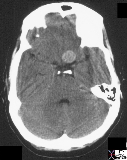
Calcified Mass
The CT scan represents a calcified aneurysm in the circle of Willis
20258 brain artery fx mass calcified dx aneurysm CTscan Courtesy Ashley Davidoff MD Uploaded RP
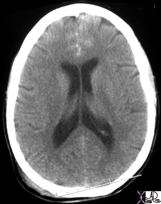
17361
17361 brain dx trauma hemorrhage Courtesy Ashley Davidoff MD Uploaded RP
|
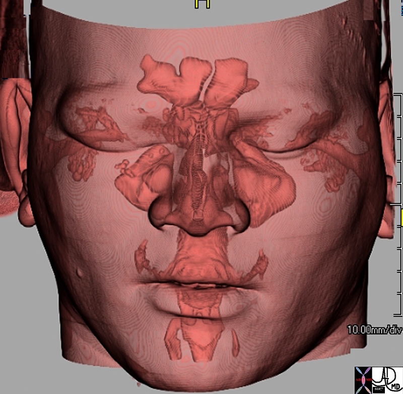
48728b01.800 |
| 48728b01.800 skull face facial bones air frontal sinus ethmoid sinus maxillary sinuses auditory canals eustachian tubes mastoid air cells nasopharynx oropharynx nasal passages sphenoid sinuses anatomy normal CTscan 3D volume rendering CTscan Davidoff MD |
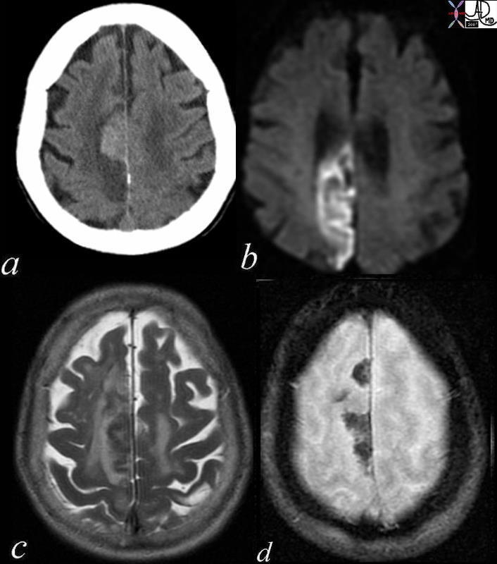
Acute and Chronic Infarction with CT and DWI MRI |
| 49679c01 brain DWI occipital lobe fx vague hypodensity right occipital lobe with encephalomalacia and ex vacuo changes in the left occipital and posterior parietal region dx acute infarction right occipital lobe chronic infarction left occipital lobe CTscan high intesity in right occipital lobe and low intensity in left occipitoparietal region dx acute infarction right occipital lobe chronic infarction left occipital lobe MRI diffusion weighted imaging Courtesy Ashley Davidoff MD |
|
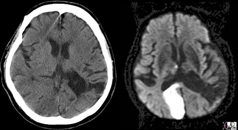
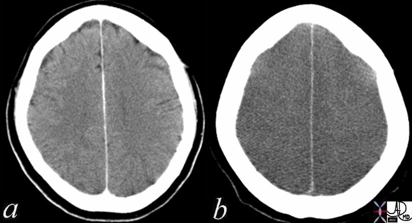
Acute Hemorrhagic Infarction of the Parietal Lobe |
| 71239c01 patient with atrial fibrillation brain cerebrum parietal lobe increased density hyperdense parasagittal hemorrhagic infarct paracentral DWII bright T2 bright GRE mixed blood products dx acute hemorrhagic infarction secondary to embolic event the patient disd have other non hemorrhagic infrctions in other parts of the brain CTscan MRI Davidoff MD |
|
Acute and Chronic by CTscan |
| 71245 brain cerebrum occipital lobe parietal lobe chronic infarction acute infarction hypodensity relative density CTscan Davidoff MD |
|
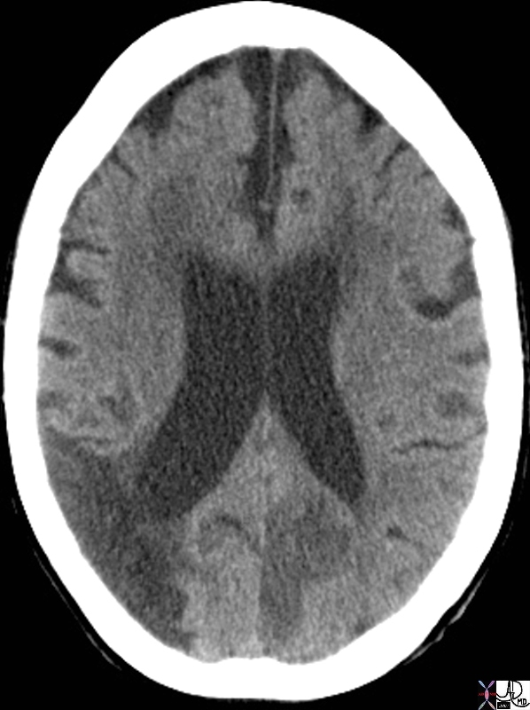
Normal – 4 months Prior and Folllowing Cardiopulmonary Arrest Loss of Gray White Differentiation |
| 70134c01 hx 52 F post cardiac arrest brain cerebrum gray matter white matter gray white differentiation gray white distinction sulci gyri loss of gray white differentiation hypodense dx anoxic injury with diffuse global cerebral edema Davidoff MD Loss of consciousness occurs within 10-15 seconds of cardio-pulmonary arrest. Irreversible brain damage can occur within 5 minutes. gray matter of the brain, particularly the frontal lobes have highest metabolic needsThe occipital, parietal, and temporal lobes and basal ganglia and cerebellum are lower. brainstem lowest needs |
Cardiovascular System
|
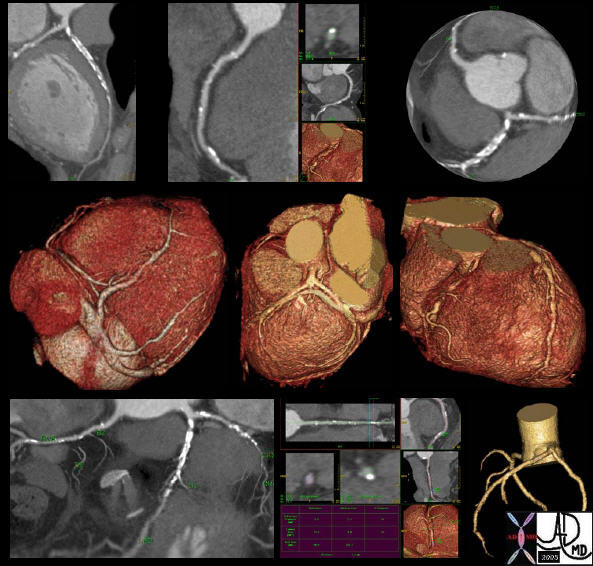
44110 |
| 44110 Case 6: Coronary Artery Disease 85-year-old male with extensive calcified plaque in the LAD and circumflex. The RCA has both hard plaque and soft plaque. Image Scan Parameters 40 x 0.625mm / 0.67mm slices / 120 kV / 699 mAs / 110mm coverage / 12 second scan Excellent vessel analysis tools include 3D Volume Rendering, MIP, color maps, and measurements. Courtesy of: Carmel Medical Center CTscan MDCT Courtesy Philips Medical heart cardiac |
|
47824 |
| 47824 heart cardiac left atrium LA left ventricle LV left atrial appendage LAA anterior leaflet of the mitral valve LV inflow LV outflow normal anatomy CTscan 3D reconstruction Davidoff MD |

|
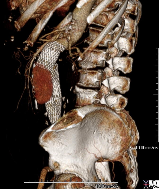
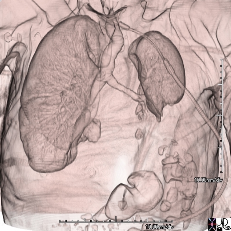
72935 |
| 72935 aorta stent graft graft leak CTscan volume rendering surface renedering treatment complication AAA 3D Courtesy Ashley Davidoff MD |
Chest and Respiratory System
|
Conventional (3-5 mm) vs High Resolution imaging (1mm) |
| 47145c02 lung interstitium interstitial disease parenchymal destruction shaggy heart border fx honeycombing interstitial pulmonary fibrosis IPF CTscan 3mm imaging and 1mm imaging – high resolution Davidoff MD |

 \ \
The Power of CTscanning of the Chest |
| 47163c01 chest lung fx ground glass pattern alveolar disease air bronchogram bronchovascular disease inflammation peribronchiole halo honeycomb interstitial fibrosis IPF CTscan Davidoff MD |

Active Tuberculosis Calcification and Air – Power of CT |
| 49812c01 50 yr F Korean fevers weight loss night sweats cough chest lung apex pleura fx calcified granulomas parenchyma pleural granulomas calcification cavitating nodules dx active tuberculosis active TB acute infection chronic infection infectious isolation Davidoff MD 49812c02 |
|
74242b01 |
| 74242b01 88 year old male emaciated thin 3D volume rendering CTscan Courtey Ashley DAvidoff MD |
Gastrointestinal System
| Air in th Bowel Wall – Ischemic Bowel |
| 16310c01.8 liver portal vein air gallbladder colon pneumatosis coli superior mesenteric vein SMV bowel ischemia ischemic bowel bowel infarction CTscan Courtesy Ashley DAvidoff MD copyright 2008 |
Methane or Nitrogen in a Cholesterol Stone |
| 70269c02 gallbladder air methane nitrogen gas cholesterol stone isodense with bile fat subtle change CTscan |
Functional Imaging

Tricuspid Regurgitation into the Hepatic Veins |
| 48098 heart cardiac liver enlarged hepatic veins fx reflux into the hepatic veins dx tricuspid regurgitation tricuspid valve congestive heart failure CHF CTscan Davidoff MD 48098c01 48098c02 48089b01 |
Musculoskeletal System
|
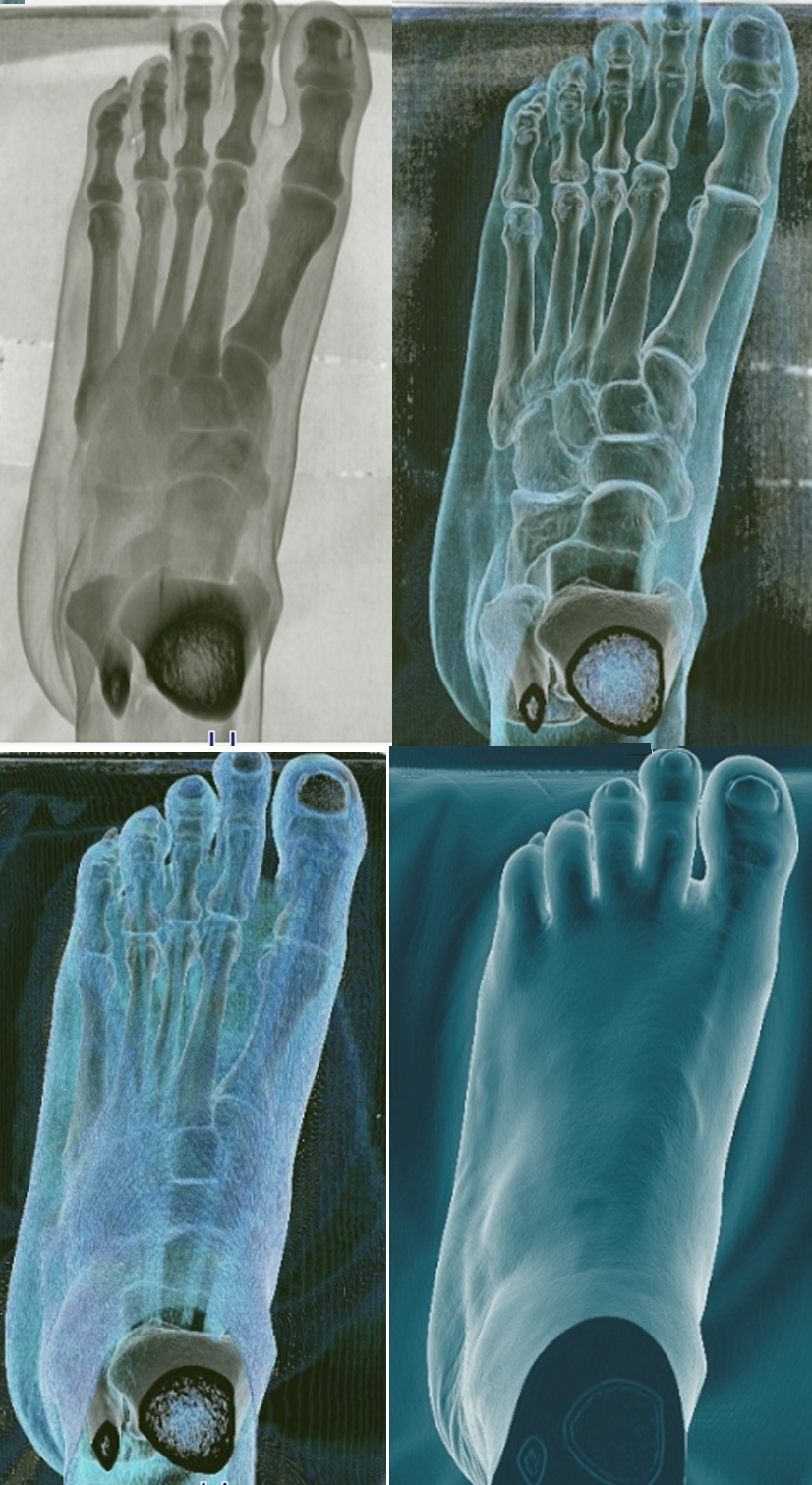
49549c01 |
| 49549c01 foot bone #D normal anatomy ligaments talus navicular cuboid tarsals metatarsal phalanges 3D volume rendering surface rendering CTscan Courtesy Ashley Davidoff MD |
|
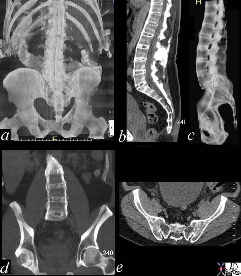
70363c03 |
| 70363c03 bone lumbar spine thoracic spine supraspinous ligament interspinous ligament abnormal bony ankylosis sacroiliac joint ankylosing spondylitis CTscan 3D Courtesy Ashley Davidoff MD |
Artifacts
Most artifacts can be attributed by the limitations of spatial resolution, temporal resolution, noise, or reconstruction algorithms.
The manifestation is reflected as blooming, blurrring, missing data.
Partial volume artifact is caused by inadequate spatial resolution as a result of voxel size that is too large, commonly seen when two structures with different density are juxtaposed, and the average of their densities is assigned to the voxels at the interface ao that their difference is not accurately reflected.. The averaging of the attenuation coefficient in a given pixel that is heterogeneous composition and the average measured in Hounsfield units is assigned to an adjacent pixel as well.