Ulcer
The Common Vein Copyright 2007
Aorta
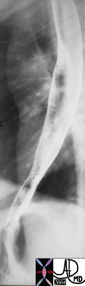
 Penetrating Ulcer with Mural Hematoma Penetrating Ulcer with Mural Hematoma |
| 47792c01 ascending aorta fx ulcer mural hematoma dx penetrating ulcer acute aortic syndrome CTscan Davidoff MD |

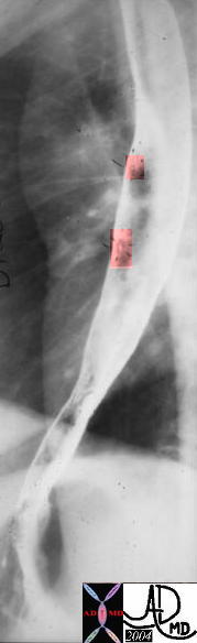
Penetrating Ulcer |
| 48363 descending thoracic aorta fx aortic ulcer fx atherosclerosis atheroma fx penetrating ulcer CTscan Courtesy Ashley Davidoff MD |
GIT
Esophagus
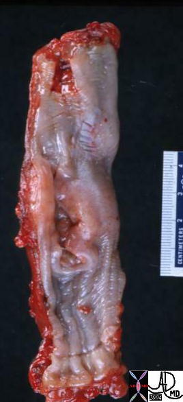
Herpes Esophagitis with Aphtous Ulcers |
| Courtesy Ashley Davidoff MD 01189 code GI esophagus + fx aphtous ulcer + dx herpes esophagitis + barium swallow upper GI UGI imaging radiology contrast X-ray infection |
The Normal (left) and Abnormal (right) Red and Swollen GE junction – Reflux Esophagitis |
| The GE junction is a distinct border as seen normally in the left image. The distal esophagus is normally a pearl white hue, while the gastric mucosa is a pink hue. In esophagitis the hyperemic inflammatory changes make the border less distinct (right image)
01239b01.800 esophagus stomach GE junction gastroesophageaal junction inflammed inflammation GERD red swollen reflux esophagitis grosspathology Courtesy Ashley Davidoff MD |
Ulcerating Squamous Cell Carcinoma of the Esophagus |
| 02425 02420 code esophagus + mass narrowing stricture ulceration heaped edges squamous cell carcinoma + grosspathology |
Normal (a) and Reflux Esophagitis (b) |
| 76217c esophagus normal smooth mucosa featurless heterogeneous mucosa nodular mucosa diffuse reflux esophagitis double contrast barium swallow Courtesy Ashley Davidoff MD |
Reflux Esophagitis with Ulcers |
| 70 year old male presents with burning substernal chest pain Barium swallow shows superficial ulceration at the GE junction and also small areas of ulceration along the distal esophagus
esophagus GE junction ulcer esophagitis barium swallow 83326.8s Courtesy Ashley Davidoff MD copyright 2009 |
Stomach
Linear Erosions of Antral Gastritis |
| 49746b01 normal
01323 01323b01 code stomach + linear erosions erosive gastritis + upper GI UGI imaging radiology contrast X-Ray inflammation |
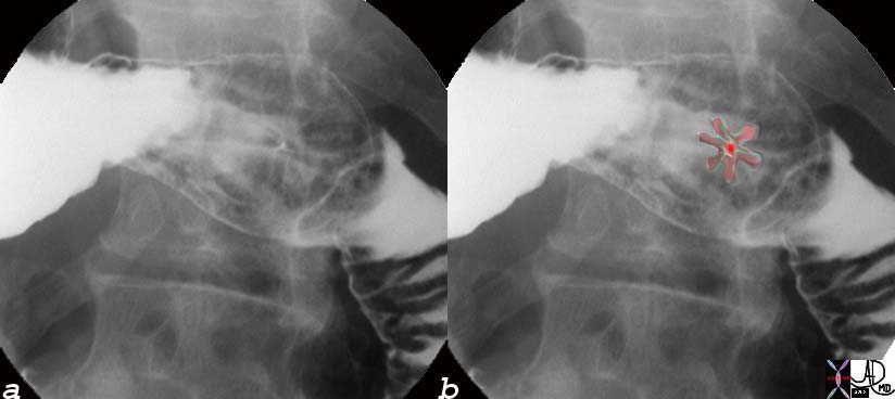
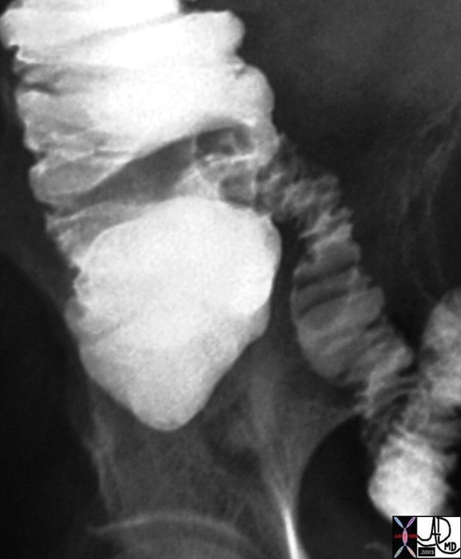
Punctate Duodenal Ulcer |
| 76121c02 duodenum bulb punctate ulcer peptic ulcer double contrast upper GI upper GI Courtesy Ashley Davidoff MD |
Small Bowel
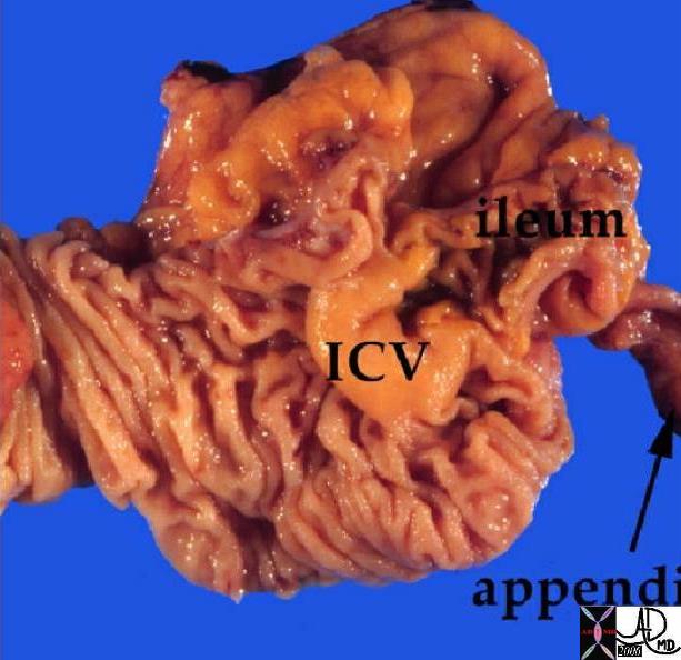 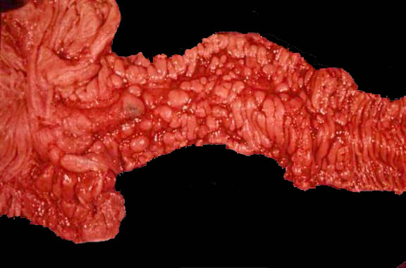
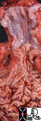
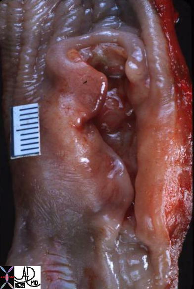
Normal and Denuded Ulcerating Mucosa with Cobblestone of the Terminal Ileum in Crohn’s Disease |
| 12231 colon large bowel cecum ileocecal valve appendix fx normal anatomy Courtesy Barbara Banner MD
02477b02 small bowel terminal ileum mucosa fx inflammation ulceration fx nodules fx cobble stone thick walled dx Crohn’s disease ileocecal valve grosspathology Courtesy Dr Gutkin MD DB |
 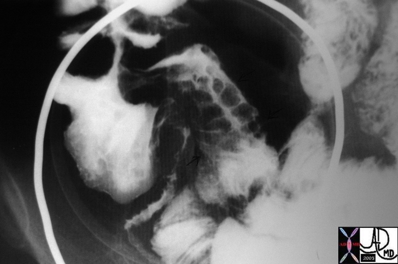
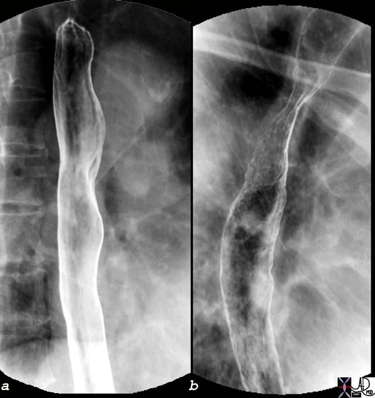
Normal and Cobblestone |
| 32525b01 small bowel terminal ileum colon ileocecal valve large bowel normal anatomy UGI SBFT Courtesy Ashley Davidoff MD
43159 small bowel fx ileum fx nodular mucosa fx cobblestone fx thickened folds fx narrowed lumen dx Crohns UGI SBFT Courtesy Ashley Davidoff MD AFD AFD
|
|
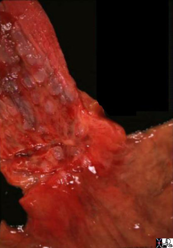
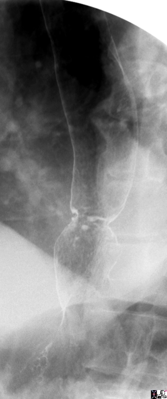
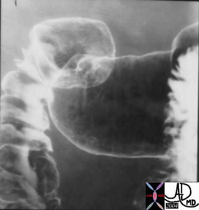
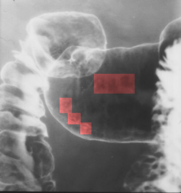
Cobblestone of the Terminal Ileum |
| 43159 small bowel fx ileum fx nodular mucosa fx cobblestone fx thickened folds fx narrowed lumen dx Crohns UGI SBFT Courtesy Ashley Davidoff MD AFD AFD
02477b02 small bowel terminal ileum mucosa fx inflammation ulceration fx nodules fx cobble stone thick walled dx Crohn’s disease ileocecal valve grosspathology Courtesy Dr Gutkin MD DB |
GUT MSK RES Skin
An aphtous ulcer is a small ulcer on a mucous membrane, usually of the mouth, characterized by an erythematous halo.