|
copyright 2007 Principles
Inflammation
Infection
Neoplasia
|


Cysts
copyright 2009
Definition
A cyst ….. is …
characterized by …..
caused by ..etiology or predisposing factors
resulting in a pathological feature (structural change or functional change) or clinical feauture
Sometimes complicated by ….
Diagnosis is suspected clinically by … and confirmed by ….
Imaging includes the use of
Treatment options depend on …. but includes …..
Etymology if available
Principles
CNS

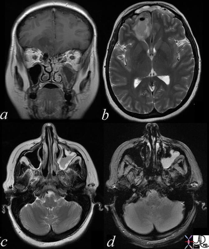
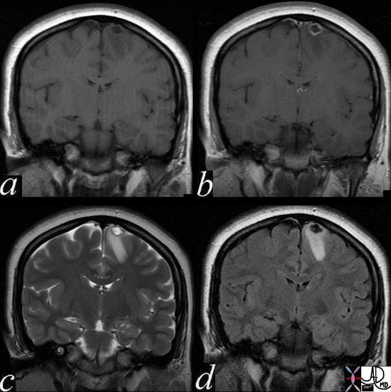
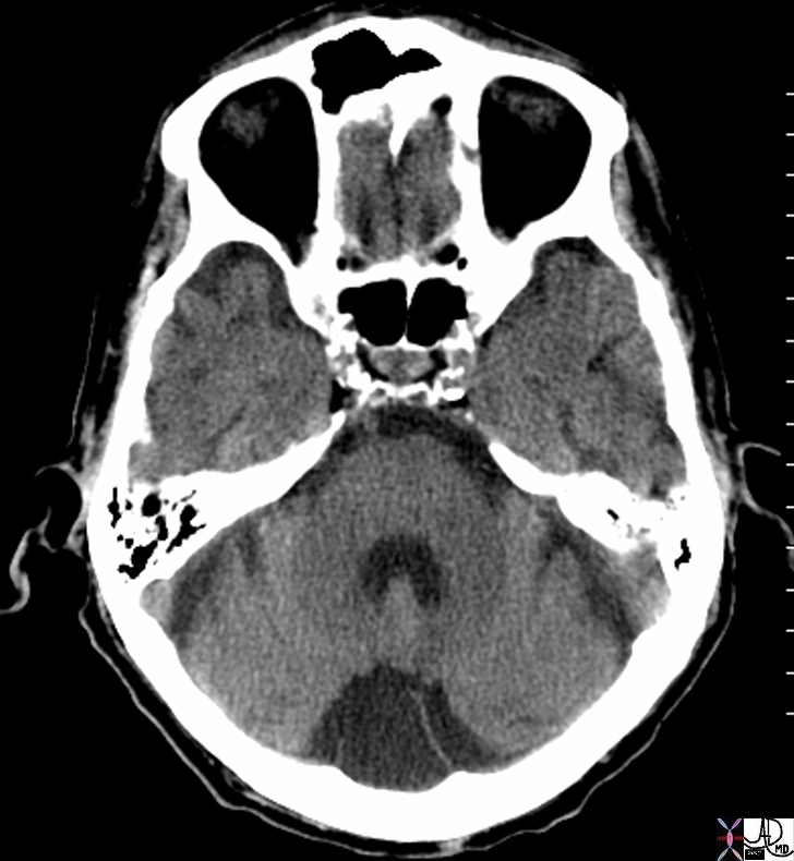
Arachnoid Cyst |
| 72179 arachnoid cyst brain meninges posterior fossa cystic collection cerebellum CSF density dx arachnoid cyst CTscan Davidoff MD |
Genitourinary Tract
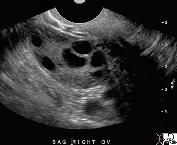
Evolving Dominant Follicle |
| 71688 ovary follicles dominant follicle normal anatomy function physiology TCV Applied Biology Cycle time USscan Davidoff MD 71689 |
Follicles in a Reproductive Female – Cyclical Phases -Size and Time |
| 71689 ovary follicles normal anatomy function physiology TCV Applied Biology Cycle time USscan Davidoff MD |

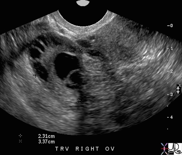
Ovulation – Mid Cycle |
| 47025c01 young patient with known ovulation one day earlier ovary Graafian follicle rupture tear drop shape pear shaped ovulation physiology normal anatomy USscan Davidoff MD |

Nabothian Cyasts in the Cervix |
| 49463 cervix fx cysts anechoic through transmission backwall enhancement dx Nabothian cysts USscan Davidoff MD |
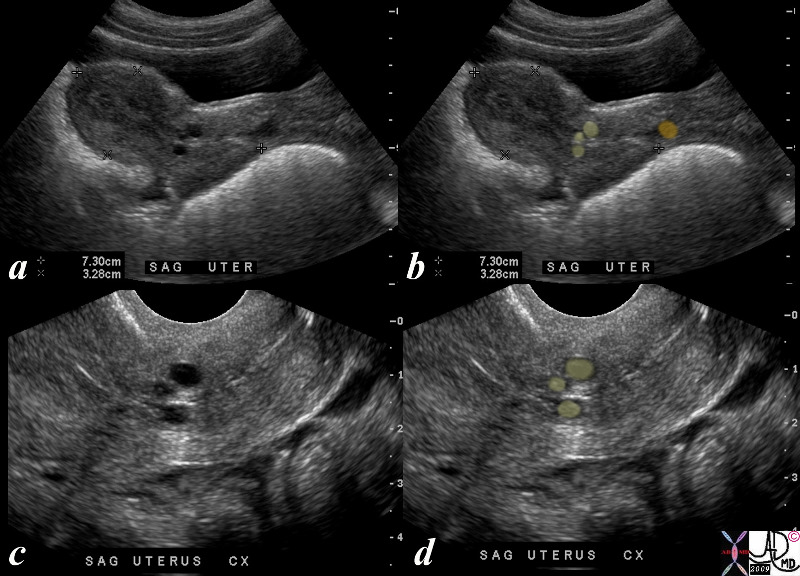
Mesonephric and Paramesonephric Cysts |
| 33year old female whose LMP was 3 weeks ago. USscan scan shows three cystic areas in the lower uterine segment (yellow overlay) likely representing mesonephric and paramesonephric cysts, and a cervical cyst likely representing a nabothian cyst.(orange) Differential diagnoses of cystic uterine lesions include cystic degeneration of uterine leiomyoma, cystic adenomyosis (adenomyotic cysts), congenital uterine cysts such as mesonephric and paramesonephric cysts, cervical nabothian cysts,
intramyometrial hydrosalpinx, and echinococcal cysts. nabothian cysts classically related to the cervix often associated with chronic cervicitis. paramesonephric or mesonephric cysts. unusual, benign congenital anomalies. not typically associated with other GU anomalies likely incidental usually followed over time rare cases of malignancy arisenin walls of these cysts. uterus lower uterine segment cysts nabothian mesonephric cyst paramesonephric cyst USscan ultrasound Courtesy Ashley Davidoff copyright 2009 all rights reserved 85453c01.8s |
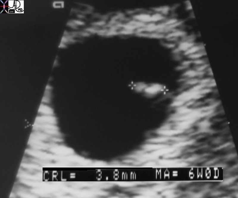
78403c02.8 |
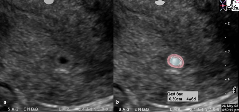
| 78403c02.8 baby pregnancy OB fetus gestational sac 4 weeks and 6 days early IUP OB endometrium no fetal pole uterus endometrium normal Courtesy Ashley DAvidoff MD |

Early Intrauterine Pregnancy |
| 27 year old female with LMP about 5 weeks ago. US shows a cystic area in the endometrial cavity consistent with an early intrauterine pregnancy. The sac measures 3.3mms consistent with a gestational age of 4 weeks and 6 days uterus endometrium
cyst 4-5 week IUP USscan ultrasound Courtesy Ashley Davidoff copyright 2009 all rights reserved 85489c01.8s |
 |

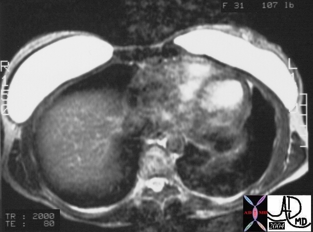
Epididymal Cyst |
| 47006 testis testes epididymis epididymal cysts through transmission backwall enhancement parts anatomy USscan Davidoff MD |
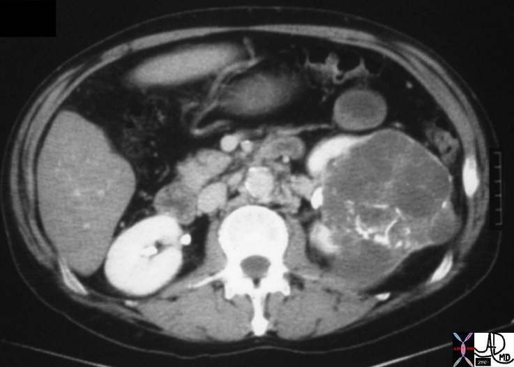
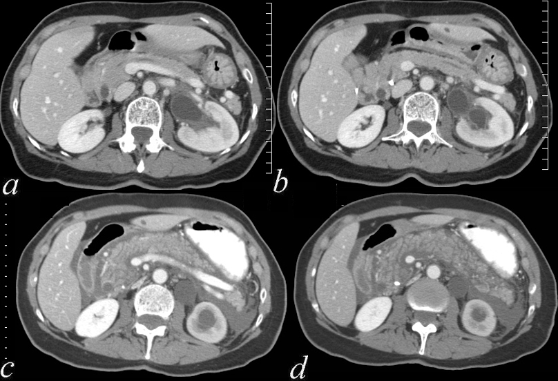
Cystic Renal Celll Carcinoma |
| 05730.800 kidney renal mass fx coarse calcifications curvilinear linear calcified space occupying displacement cystic enhancing septations dx cystic renal cell carcinoma RCC CTscan Davidoff MD Bosniak grade 4 05730.800 05730b.800 05729b |
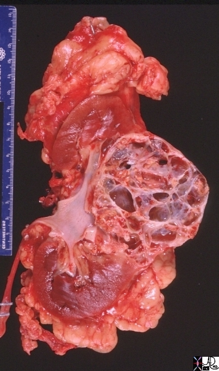
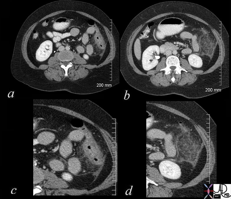
Cystic Renal Cell Carcinoma |
| 05729b kidney renal mass space occupying displacement cystic dx cystic renal cell carcinoma RCC grosspathology CTscan Davidoff MD Bosniak grade 4 05730.800 05730b.800 05729b |
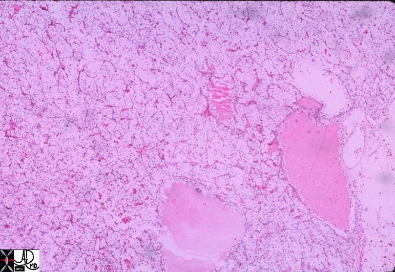
Histopathology – Cystic RCC |
| 05735 kidney renal cystic dx cystic renal cell carcinoma RCC histopathology Davidoff MD 05730.800 05730b.800 05729b 05734 |