MSK by Mina Gardezi, MS4
She has color coded things to do eg Images to get not on TCV -which did not come across on cut and paste version shown here
I will have to modify some of the images here for the IF program because the image for the IF program should be an uncluttered without overlays
| Heading | Subheading | Definition | Image | TCV link/about link | ACR appropriateness |
| dislocation | About dislocations | Dislocation occurs when the alignment of bones at a joint is disrupted, often stemming from incidents like falls, contact sports, or other high/low velocity injuries. This misalignment can result in joint instability, diminished function, and symptoms such as pain, swelling, or visible deformity. Radiographic evidence, as depicted in the listed images, showcases the misalignment of bones, with X-rays revealing larger gaps between bones than the norm. Careful examination for associated fractures on imaging is imperative, and additional soft tissue changes may prompt the need for MRI correlation to assess potential ligamentous injuries. |
|
About Knee dislocation:
https://bones.thecommonvein.net/dislocation-knee/ About Patella dislocation: |
|
| spine bilateral jumped facet | Bilateral jumped facet in the spine, also known as bilateral facet joint dislocation, represents a highly unstable injury with a notable risk of neurologic compromise. Facet joints, situated between spinal vertebrae, play a pivotal role in providing both movement and stability to the spine. This condition often arises from traumatic incidents involving buckling forces, resulting in bilateral lateral mass fractures, pedicle fractures, or locked facets. Imaging reveals the dislocation of facets on both sides of the spine, potentially leading to nerve compression, spondyloptosis, or significant neurologic deficits. |
https://prod-images-static.radiopaedia.org/images/2876762/1eacfa5f58ba848ea84320a4ad3021_big_gallery.jpg |
Pubmed article for C-spine injury: https://www.ncbi.nlm.nih.gov/pmc/articles/PMC7383727/ | ||
| subluxation | About subluxation | A subluxation is a partial dislocation where the bone doesn’t entirely detach from the joint surface, however the joint’s normal position is compromised. Traumatic injuries, ligamentous damage, chronic joint stress, or conditions like Ehlers Danlos can lead to subluxation. Physically, it is characterized by joint swelling, pain, and may or may not exhibit visible deformity. XR findings unveil partial displacement of the joint surface, abnormal joint space, and soft tissue changes, including swelling and ligamentous injuries. Chronic conditions may further manifest as bone spurs due to ongoing joint instability. |
|
||
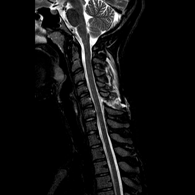
| spine anterior subluxation flexion injury | Anterior subluxation of the spine is a ligamentous disruption of the cervical spine, often stemming from an anterior flexion injury. This injury is reflected in MRI by disruption of ligaments, partial misalignment of the vertebrae anteriorly, a reversal of the typical lordotic curvature of the cervical spine, an increased gap between vertebrae, and the presence of soft tissue swelling. |
https://prod-images-static.radiopaedia.org/images/42897012/921b515e71601fc5efcff66deeacae_big_gallery.jpeg |
|||
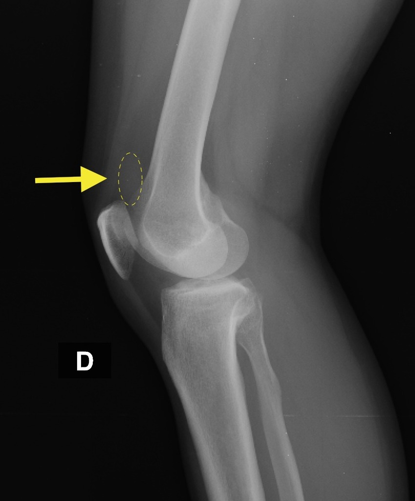
| effusion | About effusions | Effusions are an abnormal accumulation of excess fluid within a cavity, arising from a myriad of etiologies including traumatic, infectious, and neoplastic conditions. In the context of joint effusions, there is an excess accumulation of fluid in the joint cavity resulting in distension of the joint on imaging. | Hip effusion radiopedia:
https://radiopaedia.org/cases/41679/studies/44600?lang=us&referrer=%2Farticles%2Fjoint-effusion%3Flang%3Dus%23image_list_item_18118069 |
||
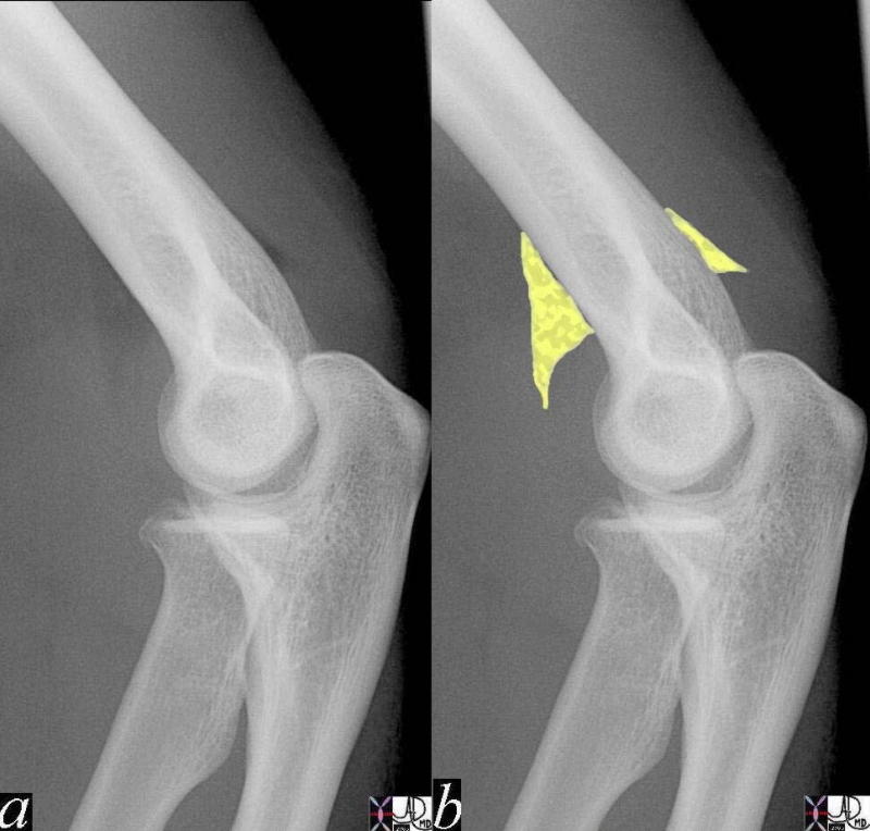
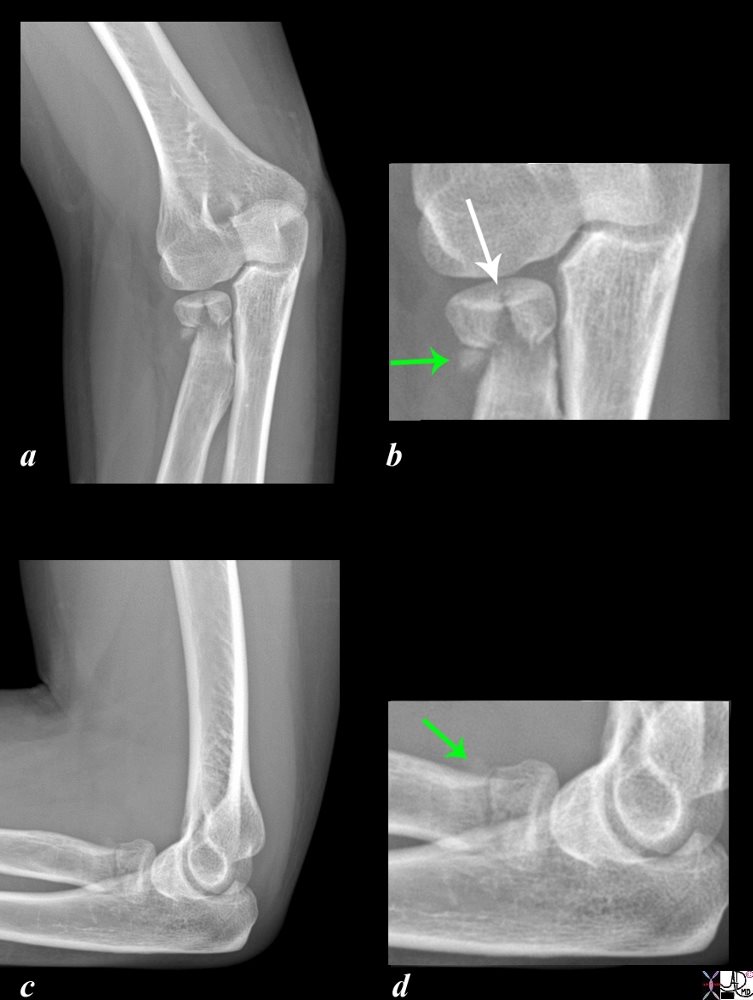
| Elbow joint effusion-radial head fracture | Elbow joint effusions caused by radial head fractures are commonly associated with a FOOSH injury (fall on outstretched hand). This type of effusion is characterized by a classic “sail sign” on XR, which is elevation of anterior and posterior fat pads caused by blood distending the joint capsule. | Sail sign:
https://beta.thecommonvein.net/wp-content/uploads/2023/05/41884c.jpg Radial head fracture:
https://beta.thecommonvein.net/wp-content/uploads/2023/03/103258c02.8s.jpg |
About radial fractures: https://beta.thecommonvein.net/fractures/radius/ | ||
| Knee effusion | In knee effusions, there is excess accumulation of fluid within the joint cavity manifests as distension and hypoechoic regions signifying fluid on ultrasound. TCauses include overuse, trauma, infections, or rheumatologic conditions like gout. While larger effusions may be detectable through physical examination alone, smaller ones necessitate imaging modalities such as ultrasound for detection. |
https://radiopaedia.org/cases/knee-joint-effusion-3?lang=us |
Knee Gout: https://bones.thecommonvein.net/gout-of-the-knee/ | ||
| Fracture Description | Introduction | Fractures are a partial or complete break in the continuity of a bone caused by trauma, overuse, or osteoporosis. There are numerous types of fractures each characterized by their respective radiographic findings. | 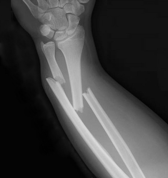
https://beta.thecommonvein.net/wp-content/uploads/2023/09/bone-0041-square.jpg |
Intro to fractures: https://fractures.thecommonvein.net/ | |
| Open (compound) | An open or compound fracture is characterized by a break in the bone that breaches the integrity of the skin leading to an external wound that can be clinically visualized. This type of fracture poses a high risk of infection, categorizing it as a medical emergency. | 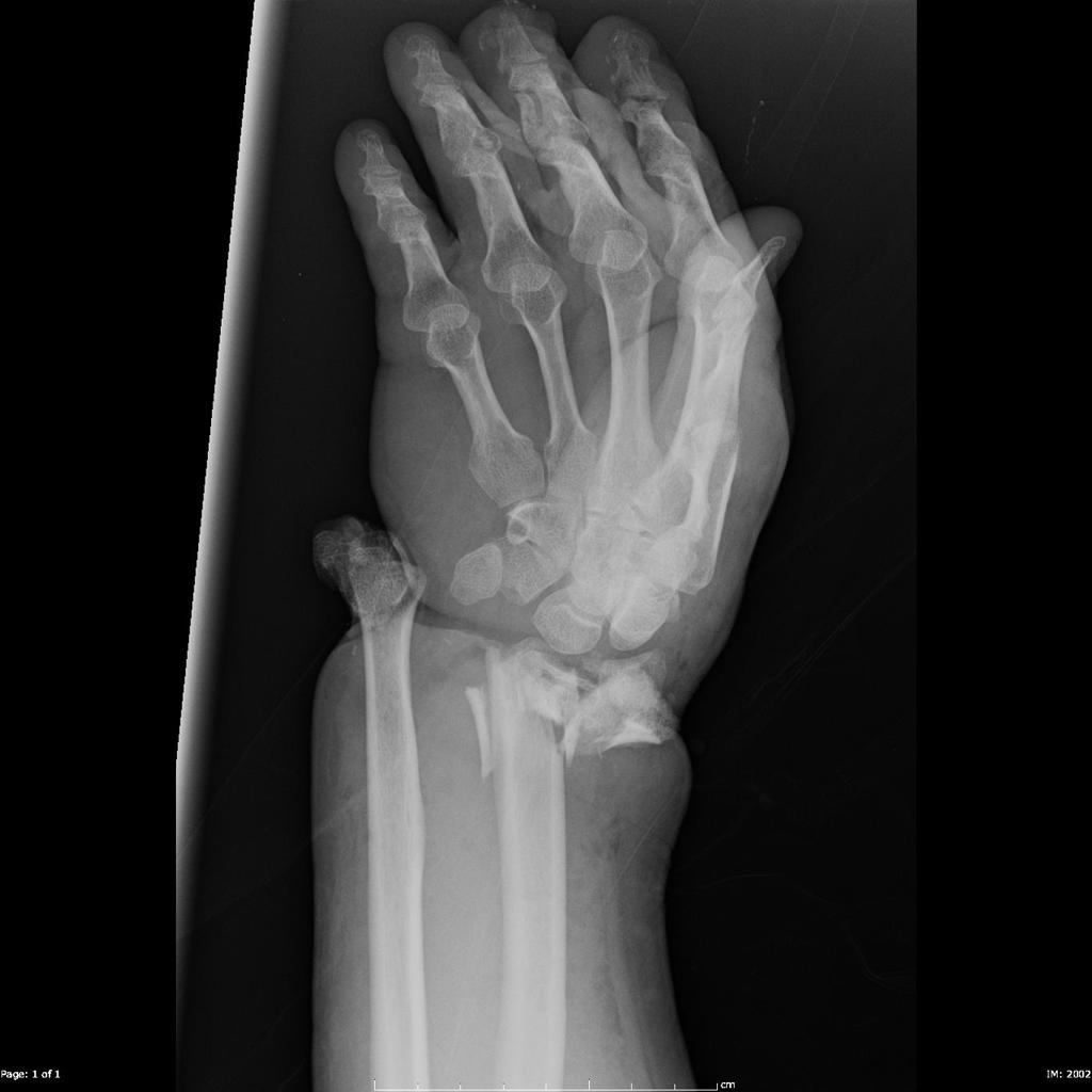
https://radiopaedia.org/cases/open-fracture-dislocation-of-the-wrist?lang=us |
|||
| Closed (simple) | A closed fracture is characterized by a break in the bone’s continuity without a break in the skin’s integrity. This is characterized by a partial or complete break in the bone seen on XR. |
https://beta.thecommonvein.net/wp-content/uploads/2023/05/72826.800.jpg |
|||
| comminuted | Comminuted fractures involve the shattering of the bone into multiple pieces, evidenced by the presence of three or more bone fragments on XR. It is a more severe fracture and carries an increased risk of delayed healing, malunion, and nonunion. |
https://beta.thecommonvein.net/wp-content/uploads/2023/03/101640b01.8.jpg |
|||
| transverse | A transverse fracture results in a break that runs horizontally perpendicular to the bone,often resulting from a high-impact collision with force applied perpendicular to the bone’s long axis. It is characterized by two distinct bone fragments on XR. |
https://beta.thecommonvein.net/wp-content/uploads/2023/03/103498fg02.8.jpg |
|||
| aligned | An aligned fracture is one where the fractured ends of the bone approximately maintain anatomical alignment. |
|
|||
| displaced | A displaced fracture is one where the fractured bone fragments shift out of anatomical alignment as visualized on XR. The degree of displacement is categorized as “mild,” “moderate,” or “severe.” |
|
|||
| impaction | In an impaction fracture, one component compresses or telescopes into another due to axial load along the bone’s long axis. It is characterized by reduced spacing between fragments, bone shortening, and a change in density in XR. |
|
|||
| distracted | Distraction fracture refers to the longitudinal separation of fracture ends, creating a gap between fragments on X-ray and resulting in an overall lengthening of the bone. |  https://beta.thecommonvein.net/wp-content/uploads/2023/03/99816c031L.81s.jpg https://beta.thecommonvein.net/wp-content/uploads/2023/03/99816c031L.81s.jpg |
|||
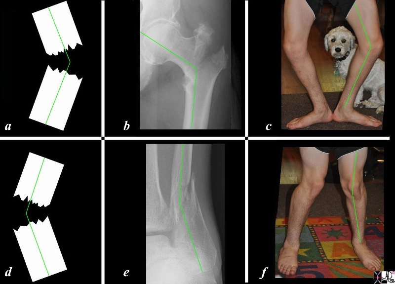
| Fracture angulation | *** | ||||
| anterior angulation | *** | Describing fracture shapes: https://beta.thecommonvein.net/fractures/shape/ | |||
| posterior angulation | *** | ||||
| medial angulation | *** | 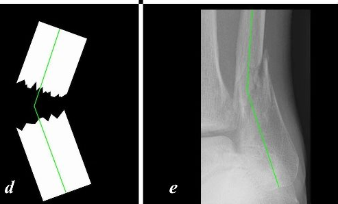
https://beta.thecommonvein.net/wp-content/uploads/2023/03/103498l02c06L.8s.jpg |
|||
| lateral angulation | *** | 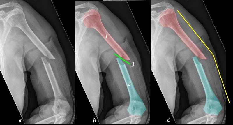 https://beta.thecommonvein.net/wp-content/uploads/2023/03/103519cd07b.8.jpg https://beta.thecommonvein.net/wp-content/uploads/2023/03/103519cd07b.8.jpg |
|||
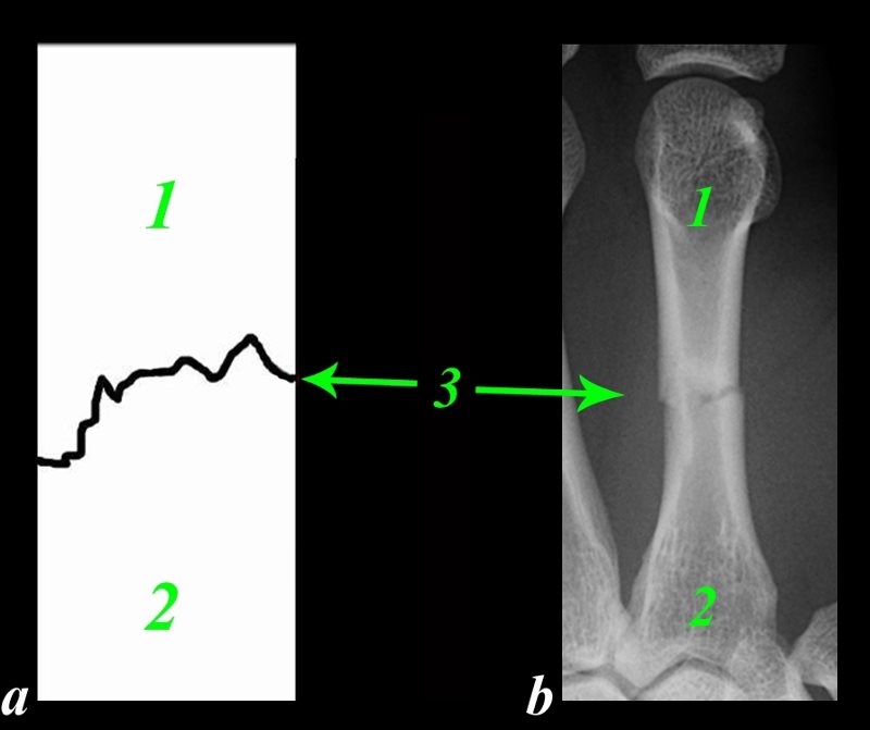
| Fracture healing | Callus | Fracture callus is part of the bone healing processoffering stabilization to the recovering bone through multiple rounds of remodeling. In the fibrocartilaginous soft callus stage, it is characterized by a subtly visible faint mass around the fracture site on X-ray, later evolving into a denser and more pronounced appearance during the hard callus stage, ultimately replaced by woven bone. |
|
||
| Bony fusion | A bony fusion is the end product of successful fracture healing wherein the previously fragmented ends reunite. It is characterized by remodeling of the hard callus, transforming it into mature compact bone and restoring a continuous bone structure as seen on XR. |
|
|||
| intra-articular involvement significance | Intra-articular involvement of a fracture signifies that the fracture line extends into the joint space as seen on XR. There may also be osseous fragments in the joint space. Intra-articular fractures are more complex than simple fractures as there is concomitant cartilage damage at the articular surface, potentially compromising joint stability and increasing risk of future posttraumatic osteoarthritis due to chondral damage. Operative intervention may be required if there are bone fragments within the joint space, which can potentially slow the healing process. | Barton’s fracture of radius with intra articular involvement:
|
|||
| Pediatric fractures | Physeal plate involvement | Involvement of the physeal plate (aka growth plate) in pediatric fractures can have significant impacts on bone growth and MSK health. Fractures at the growth plate can result in necrosis, premature closure of the growth plate, and growth disturbances, potentially leading to limb length discrepancies as other bones continue to grow unaffected. These fractures are classified using the Salter Harris classification system. |
https://beta.thecommonvein.net/wp-content/uploads/2023/03/91309c02L.8.jpg |
https://beta.thecommonvein.net/fractures/long-bones-position/ | |
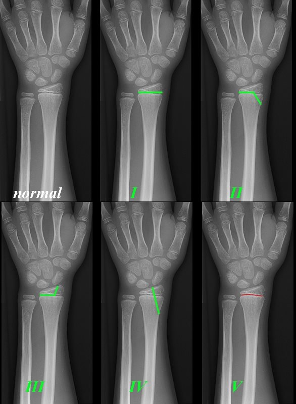
| Salter harris classification system | The Salter-Harris classification system serves as a method for grading fractures that impact the epiphyseal growth plate in pediatric cases. This classification spans from type I to type V, delineating a spectrum from less severe to more severe fractures:
|
https://beta.thecommonvein.net/wp-content/uploads/2023/03/99794bc01L.8.jpg |
https://beta.thecommonvein.net/position/bone-fractures/ | ||
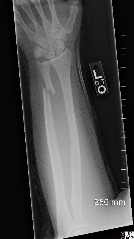
| Greenstick fracture | Greenstick fracture is a type of incomplete pediatric fracture where the bone breaks on one side while remaining intact on the opposite side. Radiographically, it is characterized by an incomplete, usually mid-diaphyseal fracture, akin to the pattern seen in a green branch that breaks incompletely on one side when bent. |
|
Fracture shapes: https://beta.thecommonvein.net/fractures/long-bones-shape/ | ||
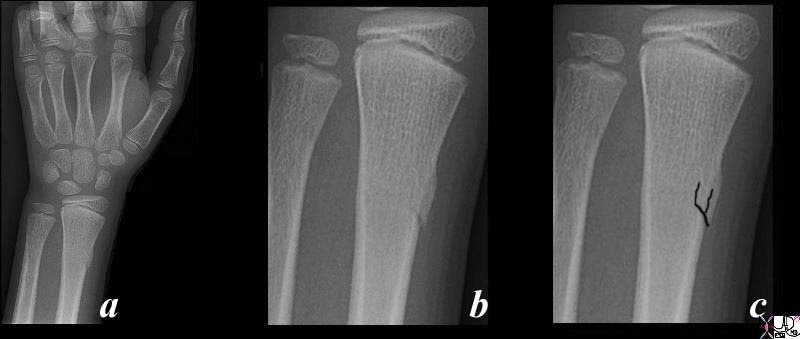
| Torus fracture | Torus fracture is a type of pediatric fracture marked by cortical buckling, presenting as a noticeable bulge on X-ray imaging without the typical appearance of fracture lines. This type of fracture is commonly situated in the metaphysis. |
|
|||
| Metaphyseal corner fractures | Metaphyseal corner fractures, colloquially known as bucket handle fractures, are defined by the fracture occurring at the metaphyseal corner, creating a fragment resembling a “bucket handle.” This fracture pattern is typically the result of twisting or pulling on a limb. While it can be associated with non-accidental trauma, particularly in children under the age of two, it is imperative to conduct thorough investigations. These injuries may also arise from normal childhood activities, necessitating careful consideration and evaluation. |
https://prod-images-static.radiopaedia.org/images/894121/9dd433fda2660a0aa4e19d1836a259_jumbo.jpg |
|||
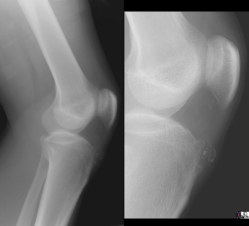
| Osgood Schlatter Disease | Osgood-Schlatter Disease emerges during adolescence, primarily as a result of knee overuse, especially in active youths engaged in activities like jumping and kicking. It is identifiable by soft tissue swelling along the patellar tendon margins, later progressing to bone fragmentation at the tibial tuberosity, as evidenced on X-rays. Managing this condition often involves activity modification and rest. Notably, Osgood-Schlatter Disease is self-limiting, typically resolving upon completion of the adolescent growth spurt. |
|
|||
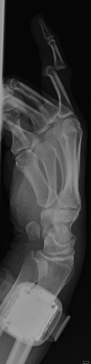
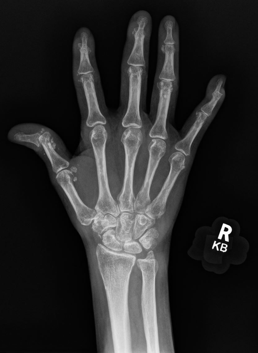
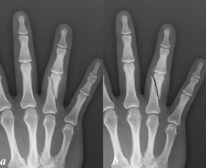
| Hand Fractures | Metacarpal/phalangeal fracture | Fractures affecting the metacarpal and phalangeal bones, commonly resulting from falls, sports injuries, or direct trauma, represent injuries to the hand. Metacarpal fractures manifest in the shaft, base, neck, or head of these bones, while phalangeal fractures exhibit variations in the proximal, middle, and distal segments of the phalanx. |
|
About phalanges fractures: https://beta.thecommonvein.net/fractures/phalanges-foot/ | |
| Boxer’s fracture | A Boxer’s fracture, colloquially termed, refers to a metacarpal neck fracture, predominantly occurring in the 5th metacarpal and visible on XR. This type of fracture is frequently attributed to impacting a rigid surface with a closed fist. Contrary to its name, it is often observed in individuals who execute punches improperly rather than professional boxers. |
|
Trauma: https://beta.thecommonvein.net/disease/trauma/
Beauty of hand: |
||
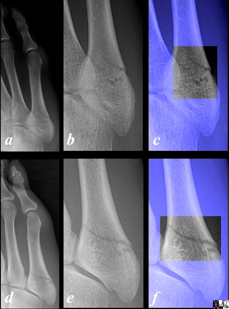
| Ankle Fractures | Ankle Malleolar fractures | Ankle fractures are classified based on the number of malleoli involved. Unimalleolar fractures typically entail the fracture of either the medial or lateral malleolus. In bimalleolar fractures, both the medial and lateral malleoli are affected, potentially disrupting the ankle mortise—the joint surface formed by the tibia and talus bones which allows for dorsiflexion and plantar flexion. Trimalleolar fractures, representing a more severe scenario, encompass fractures in all three malleoli: medial, lateral, and posterior. |
|
Fibula fractures: https://beta.thecommonvein.net/fractures/fibula/ | |
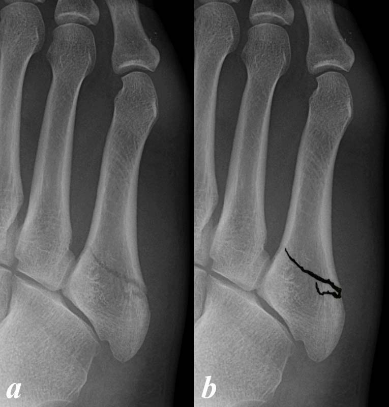
| Metatarsal fractures | Base of 5th metatarsal fracture | Fractures to the base of the fifth metatarsal are seen on xr and can be categorized based on their specific locations including tuberosity avulsion fractures, metaphyseal-diaphyseal junction fractures (aka Jones), and diaphyseal fractures. These fractures can be classified using the Stewart Classification for proximal fifth metatarsal fractures which divides them into five different categories. |
Avulsion fracture on radiopedia: https://prod-images-static.radiopaedia.org/images/10772921/077e075ee6a04271c9284f5e1303b3_jumbo.jpeg |
Stewart Classification on Radiopedia:
https://radiopaedia.org/articles/stewart-classification-for-proximal-5th-metatarsal-fractures?lang=us |
|
| Jones Fracture | A Jones Fracture, colloquially termed, refers to a proximal metaphyseal-diaphyseal fracture occurring in the 5th metacarpal and visible on XR. Due to the location of the fracture, it can be challenging to treat and has a higher risk of non-union and thus must be closely managed. |
https://beta.thecommonvein.net/wp-content/uploads/2023/05/99716b01c01.jpg |
|||
| Spine | Vertebral body anatomy on imaging? | *** | |||
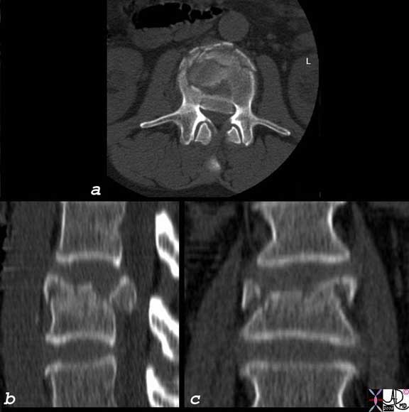
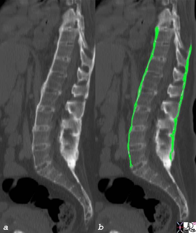
| Spinal burst fractures | A spinal burst fracture is a type of vertebral fracture characterized by a disruption in both the anterior and posterior aspects of the vertebral body. The term “burst” reflects the compression of the vertebral body in multiple directions. These fractures often result from significant axial loading, meaning along the length of the spine. Common mechanisms of injury include motor vehicle accidents, head first falls, and certain sports injuries. With burst fractures there is potential for retropulsion of a fracture fragment into the spinal cord leading to compression of the spinal cord or nerve roots as seen on CT. |
https://beta.thecommonvein.net/wp-content/uploads/2023/03/back-pain-p-044.jpg
|
Cervical spine fractures:https://beta.thecommonvein.net/fractures/cervical-spine/ | ||
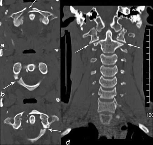
| Jefferson Fracture | A Jefferson Fracture, colloquially termed, refers to a burst fracture of the atlas bone (C1 vertebrae) which surrounds the spinal cord. This fracture is commonly caused by axial loading of compression force injuries to the head, such as those encountered in motor vehicle accidents or diving headfirst into shallow water. The distinct fracture pattern is evident on CT scans, revealing fractures in both the anterior and posterior arches of the atlas. As there is possible spinal cord involvement, Jefferson fractures need prompt diagnosis and management. | 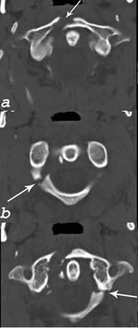 https://beta.thecommonvein.net/wp-content/uploads/2023/03/neck-pain-p-013.jpg https://beta.thecommonvein.net/wp-content/uploads/2023/03/neck-pain-p-013.jpg |
|||
| C2 fractures, dens type I-II-III | *** | ||||
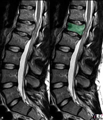
| Compression fracture- thoracic and lumbar vertebrae | A spine compression fracture involves the collapse or compression of a vertebrae, primarily occurring in the thoracic or lumbar regions of the spine. Compression fractures are typically caused by weakened vertebral bones, often due to osteoporosis or trauma. They are characterized on imaging as loss of vertebral body height due to the collapse and may be accompanied with a change in spine alignment. They are graded as mild, moderate, or severe and management is based on severity. |
https://beta.thecommonvein.net/wp-content/uploads/2023/03/back-pain-p-045.jpg |
|||
| Spinous process fracture | A fracture of the spinous process, the bony prominence extending from the vertebral arch, is typically caused by direct trauma, hyperflexion, or hyperextension of the spine. Notably, this type of fracture is considered stable and can be managed conservatively. |
|
|||
| Spondylolisthesis | Spondylolisthesis is characterized by the forward displacement or “slippage” of one vertebra over another. It can be caused by a degenerative, traumatic, or pathologic condition. Its severity is systematically graded using the Meyerding classification, which quantifies the extent of slippage. |
|
|||
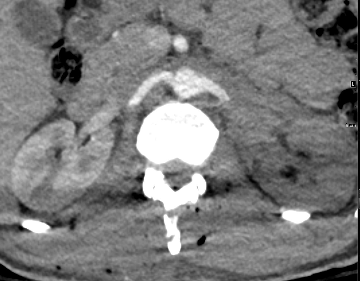
| Disc herniation | Disc herniation occurs when the soft inner core of an intervertebral disk protrudes between spinal vertebrae and can be caused by age related degeneration, trauma, lifting heavy objects, or genetic predisposition. It is characterized on MRI or CT of the spine with herniation of disc material into the spinal canal and imaging can further delineate if there is associated canal narrowing or nerve compression. |
https://beta.thecommonvein.net/wp-content/uploads/2023/03/RAM-012c.jpg |
Clinical strategies- Back pain:
https://beta.thecommonvein.net/appliedanatomy/pain-back-pain/ |
||
| Ankylosing Spondylitis | Ankylosing spondylitis (AS) is a chronic inflammatory arthritis primarily affecting the spine. AS is caused by inflammation and characterized by fusion of the vertebrae and SI joints on imaging. |
|
|||
| Upper extremity Fractures | Clavicle Fracture | Clavicle fractures typically result from traumatic incidents, such as a direct blow to the shoulder, a fall onto the shoulder, or, in some instances, a birth injury. In some cases they may lead to a visible deformity at the fracture site which is confirmed on XR. They are classified using the Allman Classification which classifies them based on location at the proximal, middle, or distal portion of the clavicle. Treatment may vary depending on the severity of the fracture, but the majority are managed conservatively through the use of a sling. |
|
About Clavicle fractures: | |
| Distal humerus fracture in a child | Distal humerus fractures, especially supracondylar ones, are predominantly observed in children and are typically the result of accidental falls. While these fractures can be identified on imaging, it’s worth noting that often the fracture line is not evident. In such cases, alternative signs, such as the “sail sign” (elevation of the anterior fat pad due to joint effusion), provide more discernible indicators of the injury. |
https://prod-images-static.radiopaedia.org/images/530232/a09b44e02b25f117743f3d11a299cf_jumbo.jpg |
|||
| Proximal humerus fracture | *** | 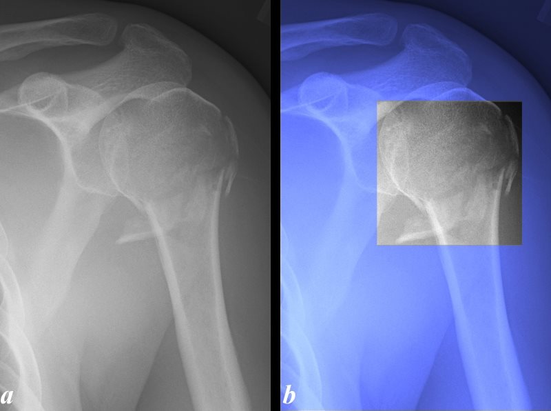 https://beta.thecommonvein.net/wp-content/uploads/2023/03/100028c.8L.jpg https://beta.thecommonvein.net/wp-content/uploads/2023/03/100028c.8L.jpg |
About Humerus fractures:
https://beta.thecommonvein.net/fractures/humerus/ |
||
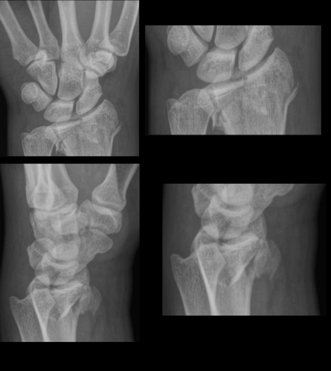
| Scaphoid fracture | *** | 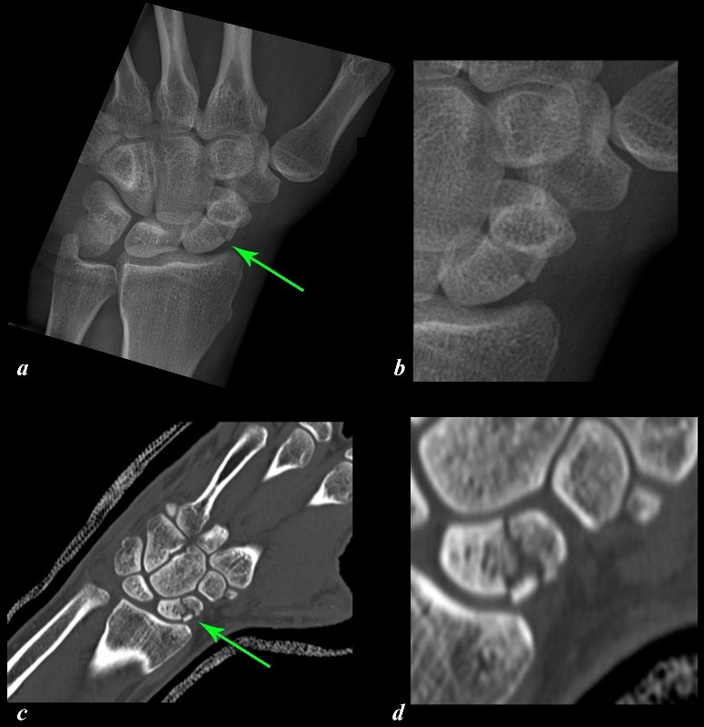 https://beta.thecommonvein.net/wp-content/uploads/2023/03/107550c01L.8.jpg https://beta.thecommonvein.net/wp-content/uploads/2023/03/107550c01L.8.jpg |
About Scaphoid fractures:
https://beta.thecommonvein.net/fractures/scaphoid/ |
||


 https://beta.thecommonvein.net/wp-content/uploads/2023/03/99718cb03L.01.8s.jpg
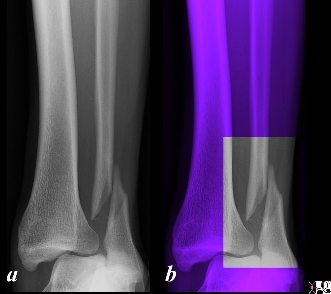 https://beta.thecommonvein.net/wp-content/uploads/2023/03/99854.6b01c001.jpg
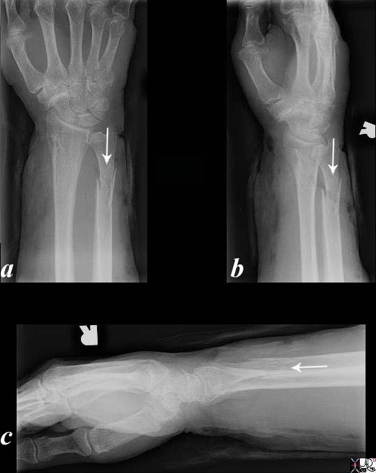 https://beta.thecommonvein.net/wp-content/uploads/2023/03/76722c.81L.jpg
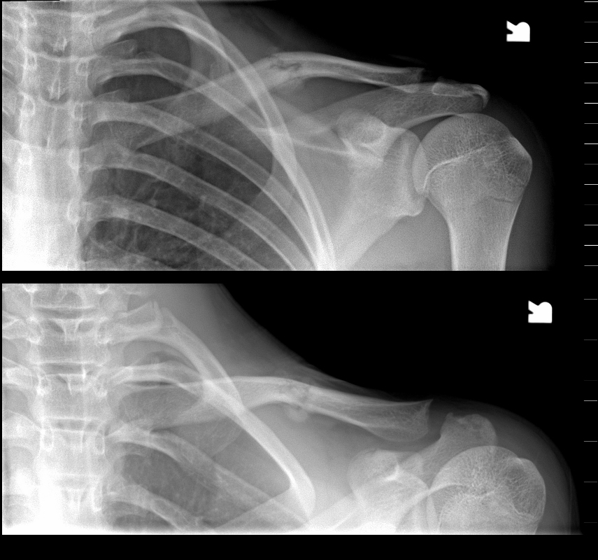 https://beta.thecommonvein.net/wp-content/uploads/2023/03/99855c.8.jpg
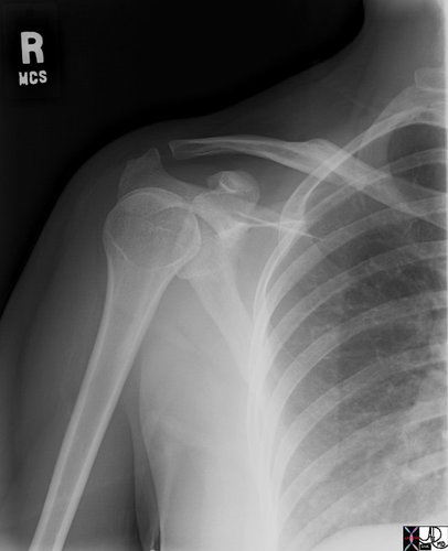
 https://beta.thecommonvein.net/wp-content/uploads/2023/03/wrist-Bartons-fracture-003-X-ray-AP-51F.jpg
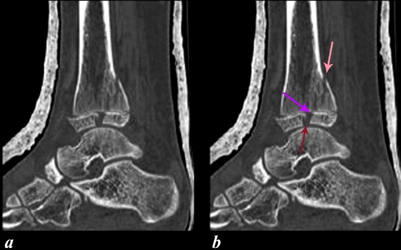
 https://beta.thecommonvein.net/wp-content/uploads/2023/08/100074c01.81L.jpg
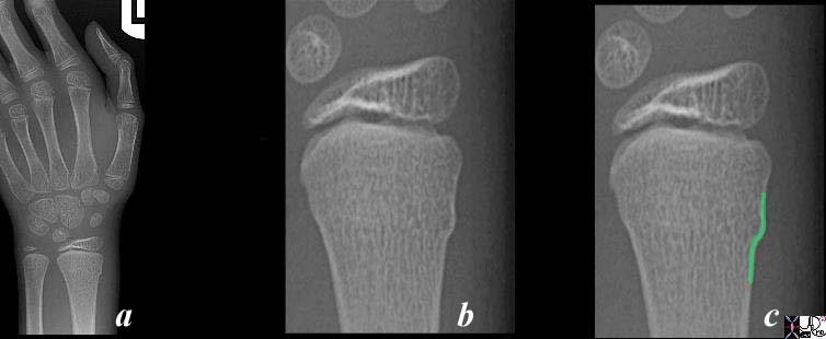 https://beta.thecommonvein.net/wp-content/uploads/2023/03/99791c01c.8L.jpg
 https://beta.thecommonvein.net/wp-content/uploads/2023/03/99941.8cL1.jpg
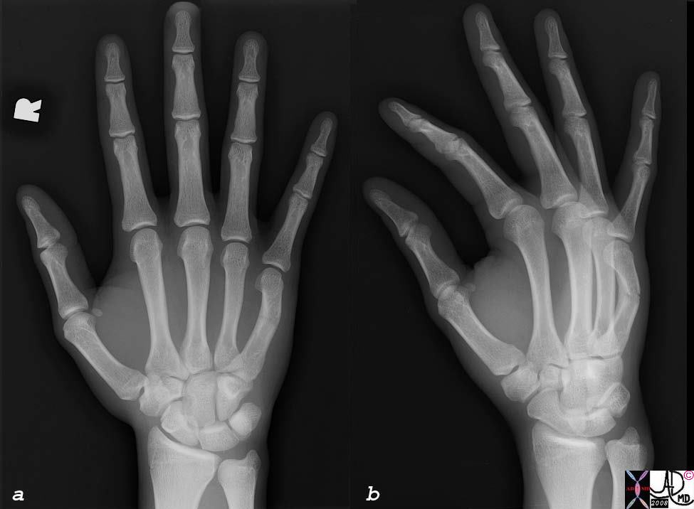 https://beta.thecommonvein.net/wp-content/uploads/2023/05/77203c01s.jpg
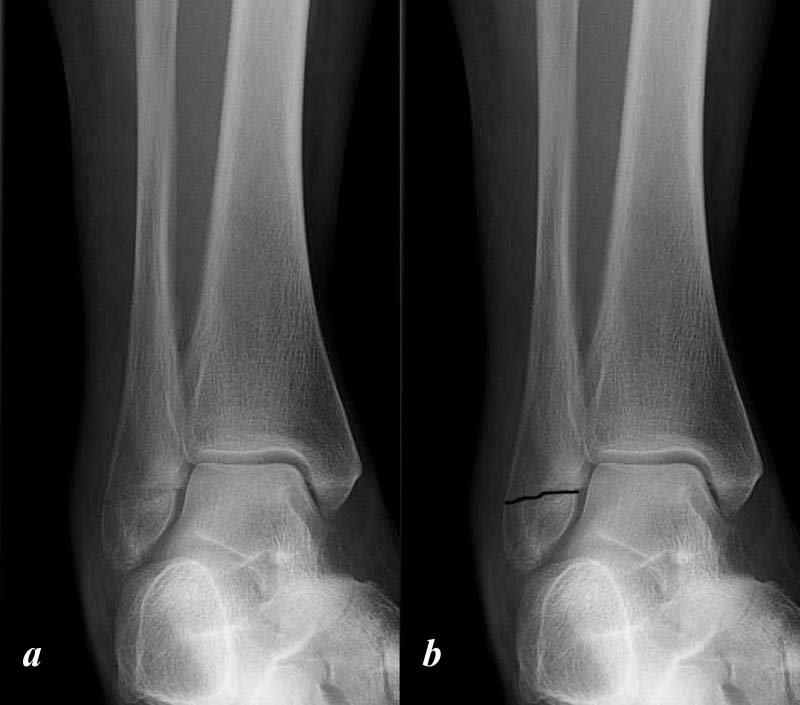 https://beta.thecommonvein.net/wp-content/uploads/2023/03/101128.8cL.jpg
 https://beta.thecommonvein.net/wp-content/uploads/2023/11/135686.kidney-trauma-Grade-V-renal-artery-occlusion-CT.jpg
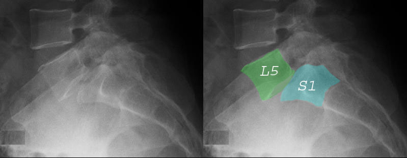 https://beta.thecommonvein.net/wp-content/uploads/2023/05/73860c03.jpg
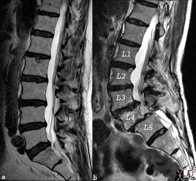 https://beta.thecommonvein.net/wp-content/uploads/2023/05/73855c01.8s.jpg
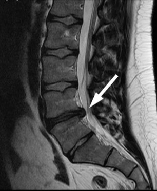
 https://beta.thecommonvein.net/wp-content/uploads/2023/05/81754c07s.jpg
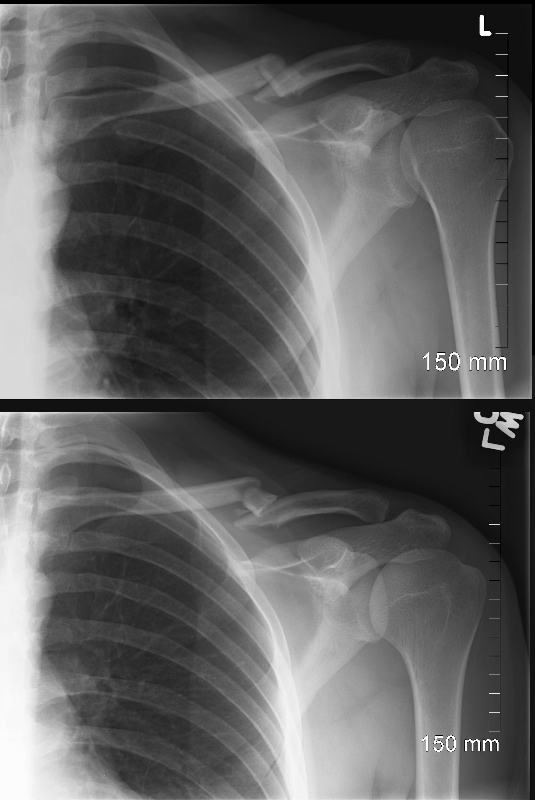 https://beta.thecommonvein.net/wp-content/uploads/2023/03/99858c.8.jpg
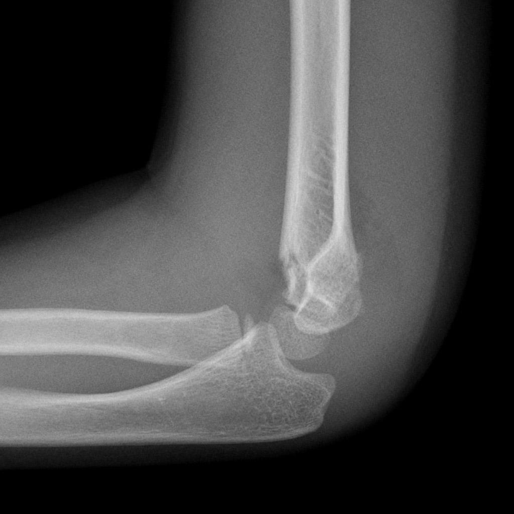
Orange= pending ortho discussion
Red= added by Mina, not on AMSER list
Yellow= No image on TCV, found on radiopedia
-69 total terms in MSK section
23 missing images on TCV
- Bilateral jumped facet in spine
- Anterior subluxation flexion injury of spine
- Effusion (any joint)
- Knee effusion
- Open/compound fracture
- Metaphyseal corner fracture- pediatric
- Toddler fracture
- Distal humerus fracture in a child
- Anterior Glenohumeral Dislocation
- Hills Sachs defect
- Pelvic ring fracture
- Tibial Plateau fracture
- Base of 5th metatarsal fracture (not jones)
- Lisfranc fracture-dislocation
- C2 fractures, dens type I-II-III
- Spondylosis
- Benign bone mass
- Disuse osteopenia
- Ligament injury
- Spine posterior ligamentous injury
- Knee ligament Injury-ACL tear
- Knee meniscus injury-meniscus tear
- Rotator cuff injury
- Findings
-
Subject Headings for the Findings Section in MSK (Mina)dislocationsubluxationdislocation spine bilateral jumped facetssubluxation spine anterior subluxation flexion injuryeffusionseffusion elbow joint , radial head fractureeffusion kneefracturesintroduction see fractures.thecommonvein.net)fractures open (compound)fractures closedfracture simplefracture comminutedfracture transversefracture alignedfracture displacedfracture impactionfracture distractedfracture anterior angulationfracture posterior angulationfracture medial angulationfracture lateral angulationfracture healing callousfracture healing bony fusionfracture intra-articular involvement significancefracture pediatric significance of physeal plate involvementfracture abusefracture abuse metaphyseal corner fractures (bucket handle) in child abuse/nonaccidental traumafracture metacarpal/phalangeal fracturesfracture ankle medial and lateral malleolar fractures, mortisefracture base of 5th metatarsal fracture (NOT “Jones”!!)fracture C1 burst (Jefferson) fracturefracture claviclefracture distal humeral in a child (signs elbow effusion)fracture pediatric tibia toddler fracture tibia (get BOTH views!)fracture proximal humerusfracture radial head (signs elbow effusion)fracture scaphoid : caveats, physical exam, delayed healing; viewsfracture spine burst (mechanism of injury; significance of canal narrowing)fracture spine C2 fractures, dens type I-II-IIIfracture spine compression thoracic and lumbar vertebraefracture spine spinous processfracture tarsal-metatarsal (Lisfranc) fracture/dislocationfracture tibia plateaufracture with extension into jointfracture wrist: fall on outstretched handfracture dislocationfracture dislocation anterior gleno-humeral dislocation and Hill Sachs fracturemassmass aggressive featuresmass benign featuresosteoporosisosteopeniaosteopenia disuseosteopenia age relatedinjury ligaments tendons cartilagesoft tissue injury spine posterior ligamentous injury (subtle signs of)soft tissue injury knee ligament and meniscussoft tissue injury rotator cuff injurysoft tissue joint spine spondylosis/degenerative change vs. arthritis in spinal column
- Diseases
- introduction common sites
- arthritis inflammatory
- arthritis osteoarthritis
- arthritis septic
- Enchondroma
- metastasis blastic appropriate imaging study to assess
- metastasis lytic
- myeloma- imaging caveats
- osteomyelitis
- osteoporosis
- septic joint
- sports/overuse/stress injuries