Copyright 2008
Bronchi
|
Tram Tracking – Acute Bronchitis |
| 75715c02 young female with a cough fx tram track ines RLL right lower lobe bronchus acute bronchitis CXR plain X-ray Courtesy Ashley Davidoff MD |
 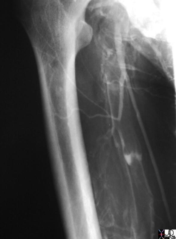
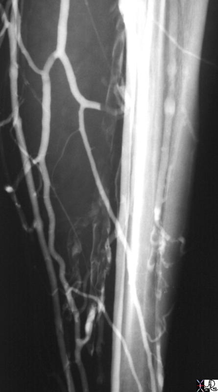
Tram tracking Acute DVT |
| 26058 26060.800 superficial femoral vein vein vein filling defect tram tracking DVT deep vein thrombosis distended venogram venography Courtesy Ashley Davidoff MD |

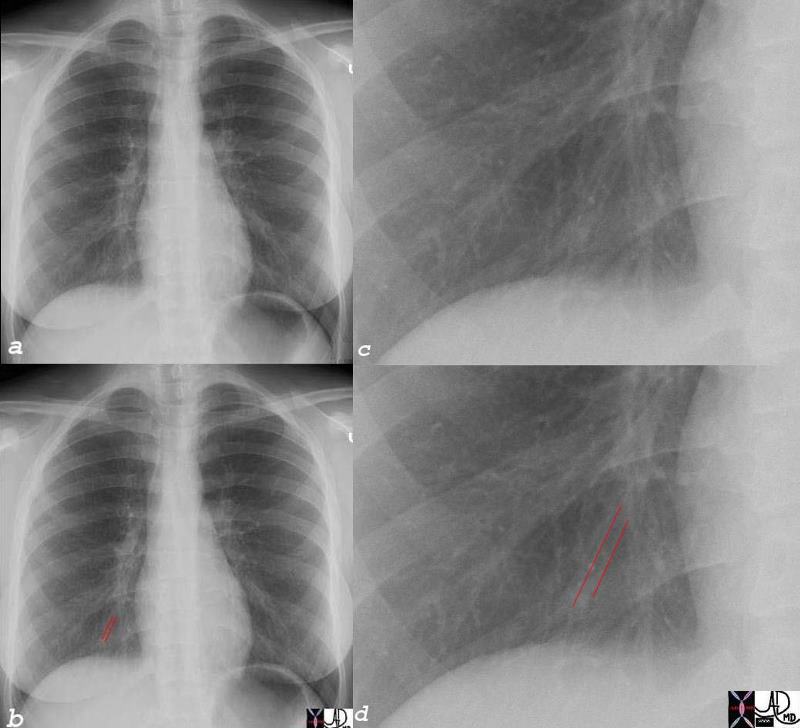
Deep Venous Thrombosis with Overlays to Demonstrate Clot and Tram Tracking |
| The venogram used to be the study of choice to rule out deep venous thrombosis until ultrasound was introduced in the 1990’s. Venography for DVT is rarely used today. However this study shows in a graphic manner, the presence of clots in the deep veins of the calf and shows the tram tracking resulting from the contrast sliding up the sides of the thrombus implying that the clot is not attached to the walls, is loose, and is therefore easily detachable into the circulation with potentially devastating results. The clot is shown overlaid in maroon in b and d. Once the clot is recognized, it is easier to go back to images a and c to identify the white column of contrast alongside the thrombus which gives the appearance of the tram-track.
code veins deep veins calf thrombosis tram tracking venogram venography Courtesy Ashley Davidoff MD copyright 2009 all rights reserved 26058c01b04.8s |
|
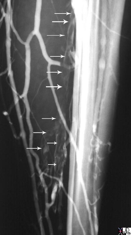
Using Arrows to Demonstrate the Tram Tracking |
|
The venogram demonstrates classical tram tracking caused by the thrombus filled calf veins with tracking of the contrast alongside the thrombus. 26058.801 vein calf vein filling defect DVT deep vein thrombosis distended venogram venography tibial veins Courtesy Ashley Davidoff MD |