Calcification
The Common Vein Copyright 2007
Ashley Davidoff MD
 CT vs MRI CT vs MRI |
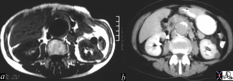
| These two images represent cross sectional imaging of an infrarenal fusiform abdominal aortic aneurysm, with image a, being from a T1 weighted MRI sequence and image b from a CT scan with contrast. MRI fails to have the sensitivity for calcium that CT does, and since calcification is an intrinsic part of aneurysmal disease, when it comes to meausurement of aortic aneurysmal size, CT is superior. Courtesy Ashley Davidoff MD 15153c code CVS aorta abdomen AAA aneurysm fusiform infrarenal |
Head and Neck
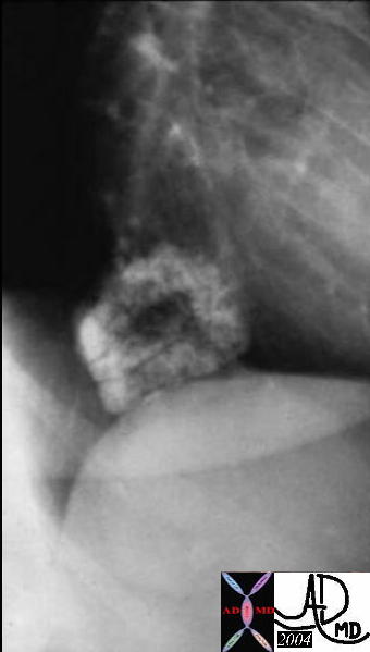
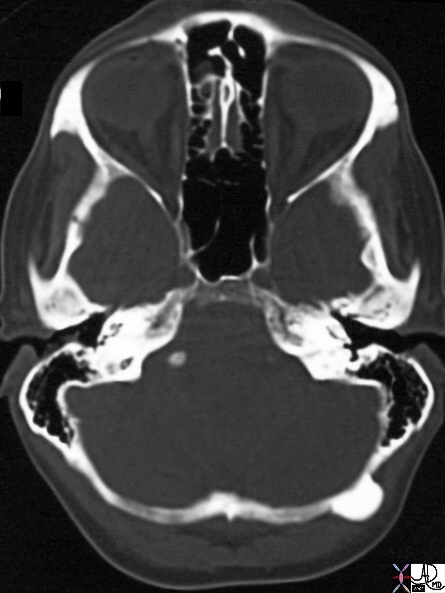 Osteoma Osteoma |
| 29107 44F one occiput occipital bone fx mass ossified dx osteoma neoplasm benign CTscan Davidoff MD |
Osteoma
Imaging findings
Size – usually less than 2 cm in size
Shape- looks like half a billiard ball
Position – Mist frequent in the frontal and ethmoid sinuses
Well-circumscribed, sharply-marginated round and
outer table of the skull
Seen in the auditory canals of swimmers and divers
Character – very dense lesions of dense compact bone
Associated findings – multiplicity – Gardners
Notes
Most common tumor of the paranasal sinuses
Benign tumor of membranous bone consisting of dense, compact bone
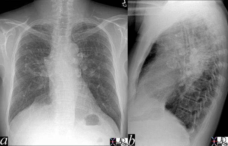
 Metastattic Calcification in Hypeparathyroidism Metastattic Calcification in Hypeparathyroidism |
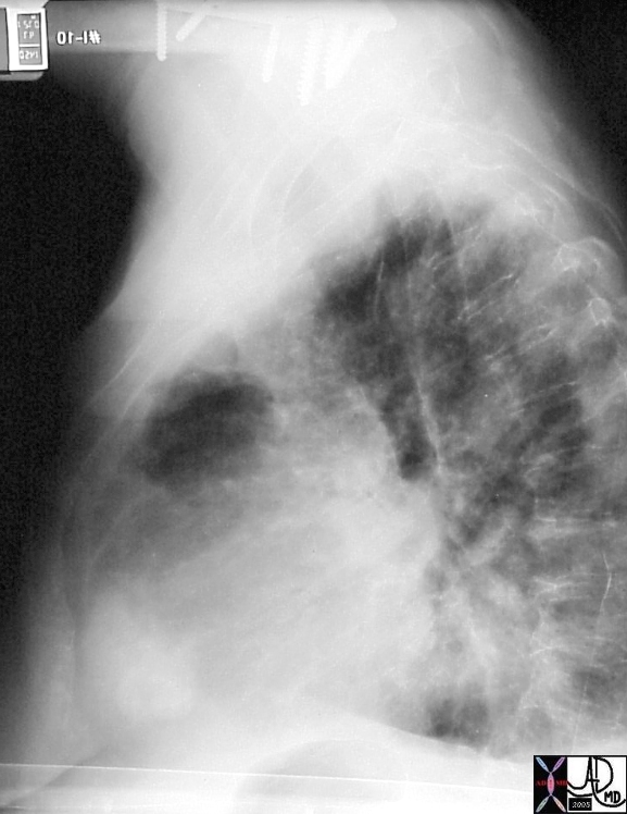
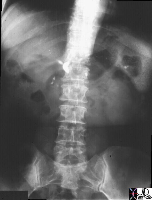
| The plain film of the chest is from a 31 year old female with hyperparathyroidism and shows clumped calcifications in the right axilla consistent with metatstattic calcification of soft tissues. The presence of the prosthesis and the asymmetry of the accumulation suggests that the trauma of surgery needs tobe implicated in the pathogenesis. The surgical clips in the mediastinum seen better on the zoomed image on the right reflects surgical footprints of a parathyroidectomy.
chest muscle calcification calcified Courtesy Ashley DAvidoff MD copyright 2009 all rights reserved chest axilla fx calcification dx dystrophic calcification dx hyperparathyroidism plainfilm 24656c.8s |
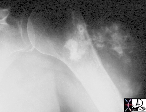 15537b07 15537b07 |
| 15537b07 hx 75F with left shoulder pain heart left humerus left shoulder fx cartilagenous popcorn type calcification dx chondrosarcoma chondrogenic sarcoma of the shoulder with metastasis to the RV right ventricle malignant imaging radiology plain film Courtesy Ashley Davidoff MD |
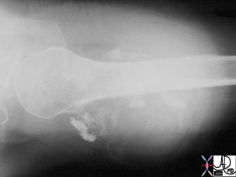
15537b08 |
| 15537b08 hx 75F with left shoulder pain heart left humerus left shoulder fx cartilagenous popcorn type calcification dx chondrosarcoma chondrogenic sarcoma of the shoulder with metastasis to the RV right ventricle malignant imaging radiology plain film Courtesy Ashley Davidoff MD |
42195c01 |
| 42195c01 Courtesy Ashley Davidoff MD medical students code calcification eggshell calcification hila hilum lung lymph node mediastinum saracoidosis |
| 33472b01.800 |
| 33472b01.800 chest lung lymph node mediastium mediastinal intraparenchymal lymp node fx eggshell calcificaton dx probable sarcoidosis CTscan Davidoff MD |
29125c |
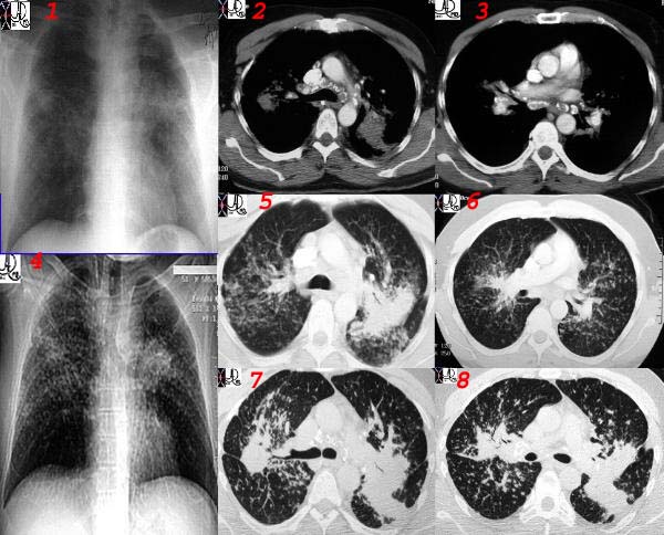
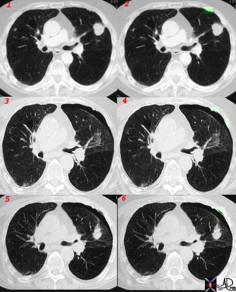
| This collage of a plain film and a CT of sarcoidosis characterised by confluent fibrosis and characteristic egg- shell calcification. Note the upper lobe predominance. Courtesy Ashley Davidoff MD. 29125 |
+
30252 |
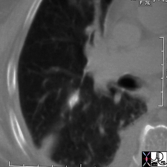
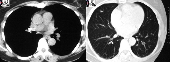
| This CT shows a prominent calcification in the right middle lobe, consistent with granulomatous disease, either TB or histoplasmosis. Note however that the calcification is eccentric and thus it is essential to follow this up on the 3/6/12month/1 year protocol. The presence of a calcified ipsilateral hilar node in this case makes granulomatous disease more likely. (30249 30252 30252c) |
31491 |
| 31491 lung fx mass lung nodule fx calcified calcifications dx rectal carcinoma complicated by pulmonary metastasis CTscan Courtesy Ashley DAvidoff MD |
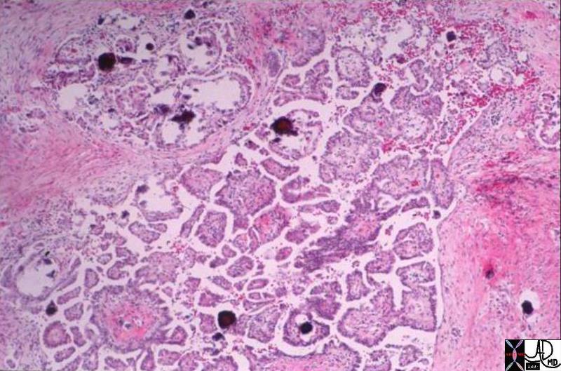
Psammomatous Calcification – Ovarian Carcinoma |
| 03779.800 liver metatstasis primary ovarian cancer adenocarcinoma psammomatous calcifications psammoma bodies glandular malignancy histopathology Davidoff MD |
30325 |
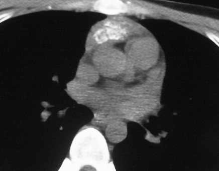
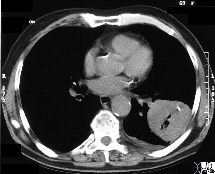
| This CT demonstrates a calcified anterior mediastinal mass. The mass proved to be lymphoma. Calcification of lymphoma prior to therapy is quite unusual. Courtesy Ashley Davidoff MD. (30321 30325) |
32017c |
| This combination image is a CT through a mass in the LUL showing focal pleural calcification overlayed in green (2) in this patient with asbestos related disease. A pneumothorax followed the biopsy revealing that the pleural plaques are positioned on the parietal pleura.(3,4) A small amount of air introduced from the xylocaine injection outlines the parietal pleura.(5,6) Courtesy Jorge Medina MD. 32017c.jpg |
01313 |
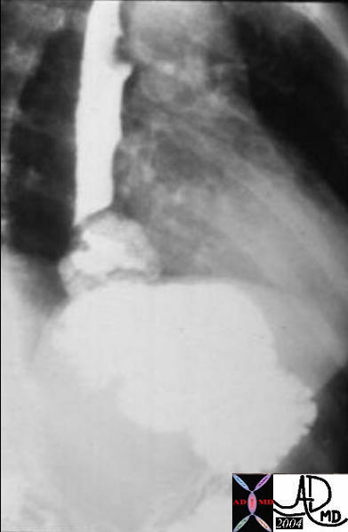
| Courtesy Barbara Banner MD 01313 code esophagus mass calcified calcification leiomyoma imaging radiology contrast X-Ray plain film CXR chest |
| 01312 |
| Courtesy Barbara Banner MD 01312 code esophagus mass calcified calcification leiomyoma barium swallow upper GI UGI imaging radiology contrast X-Ray |
| 01314 |
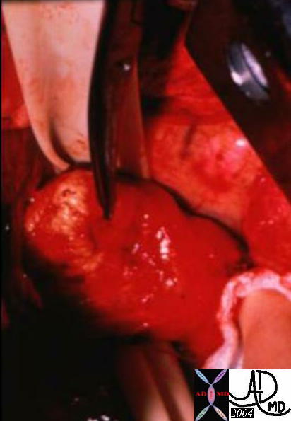
| Courtesy Barbara Banner MD 01314code esophagus mass calcified calcification leiomyoma grosspathology |
| 01316 |
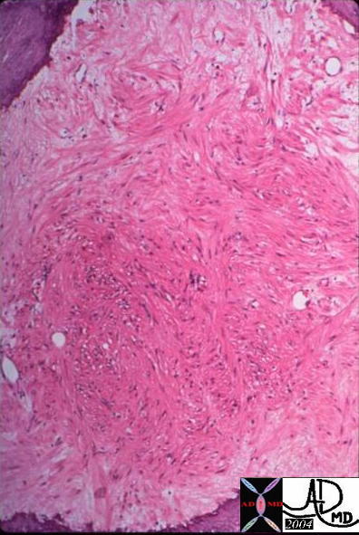
| Courtesy Barbara Banner MD 01316 esophagus mass calcified calcification leiomyoma histopathology |
| 00644 |
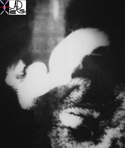
| 00644 stomach gastric fx calcified mass calcification dx leiomyosarcoma upper GI UGI imaging radiology contrast X-Ray Courtesy Ashley Davidoff MD |
| 00645 |
| 00645 stomach gastric fx calcified mass calcification dx leiomyosarcoma imaging radiology CTscan Courtesy Ashley Davidoff MD |
| 00645b |
| 00645b stomach gastric fx calcified mass calcification dx leiomyosarcoma imaging radiology CTscan Courtesy Ashley Davidoff MD |
Abdominal Cavity
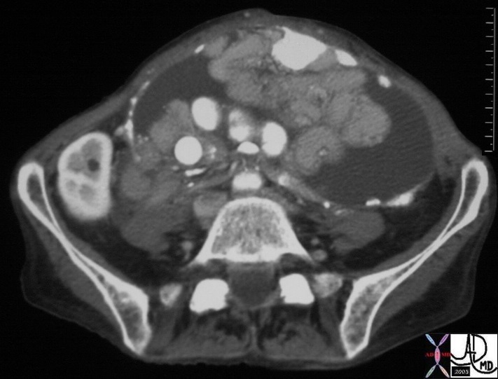
| 30368 |
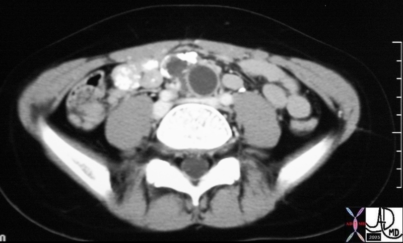
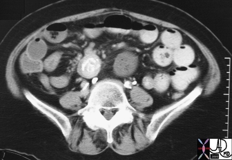
| 30368 small bowel fx encased abdomen peritoneal space peritoneum fx calcification loculated ascites dx ovarian carcinoma complicated transperitoneal metastasis CTscan Courtesy Ashley Davidoff MD DB |
| 30370 |
| 30370 small bowel fx encased abdomen peritoneal space peritoneum fx calcification loculated ascites dx ovarian carcinoma complicated transperitoneal metastasis CTscan Courtesy Ashley Davidoff MD DB |
30252 c |
| This CT shows a prominent calcified node in the right hilum, and a calcification in the RML consistent with granulomatous disease, either TB or histoplasmosis. In this case the calcification in the lung is eccentric and hence it has to be closely followed, although given the calcified hilar node malignancy is extremel unlikely. (30249 30252 30252c) code lung pulmonary nodule calcified eccentric calcification lymph node hilum hilar granuloma granulomatous disease imaging radiology CTscan |
|
| This is a CT scan show a cavitating mass in the superior segment of the left lower lobe, that also has an eccentric calcification. This is a classical example of squamous cell carcinoma of the lung. The calcification is unusual. Courtesy Ashley Davidoff MD. code lung pulmonary mass calcification eccentric cavitating cavitary neoplasm malignant primary malignancy carcinoma imaging radiology CTscan cavitation |
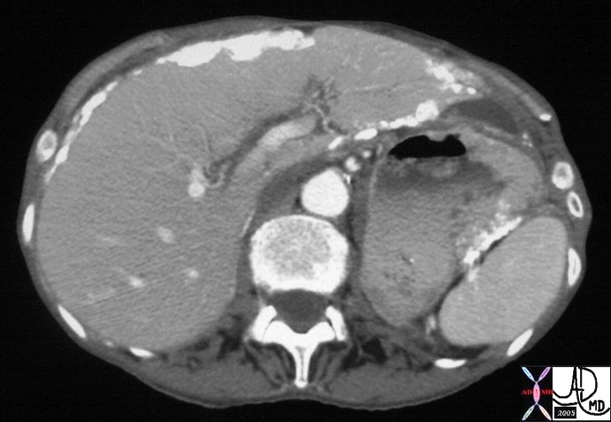
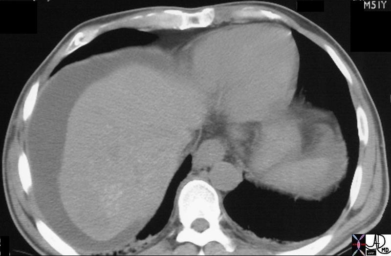
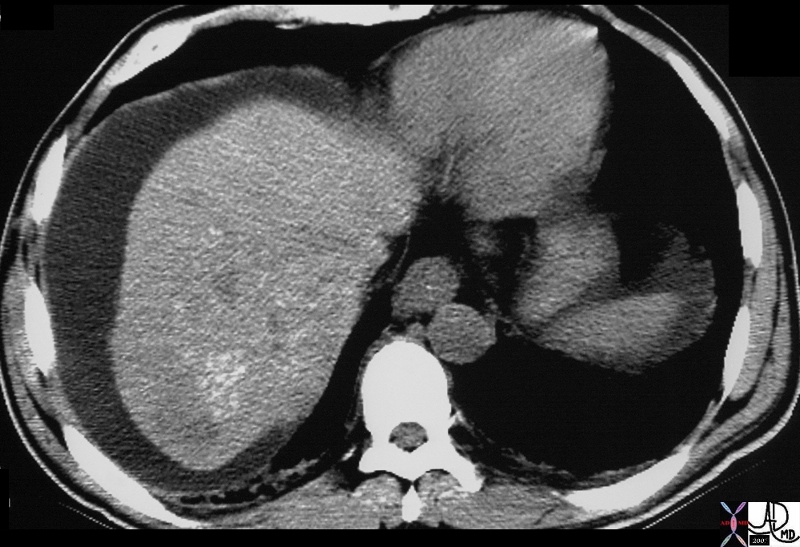
 22992 22992 |
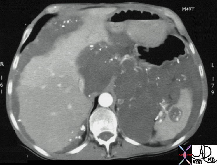
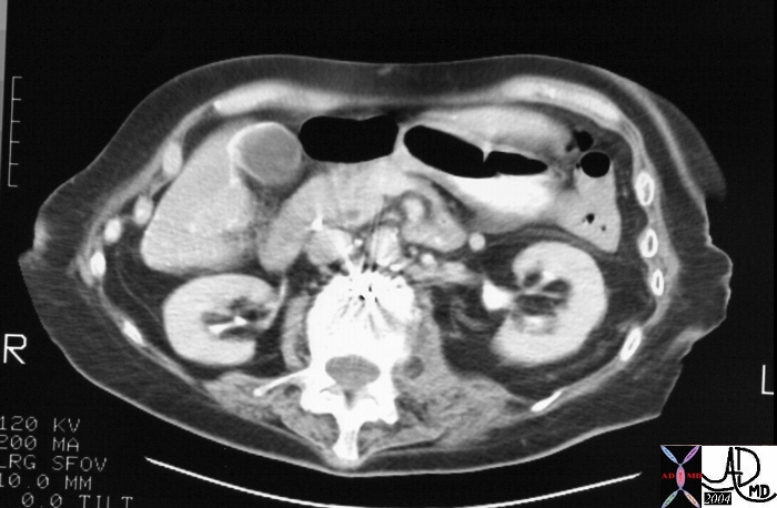
| 22992 liver + capsule + spleen small bowel stomach abdomen peritoneum peritoneal cavity fx mass + fx calcification + dx pseudomyxoma peritonei + imaging radiology CTscan Courtesy of Ashley Davidoff MD DB |
Gastrointestinal Tract
 22913 22913 |
| 22913 jejenum fx mass fx cystic fx calcification dx small bowel duplication imaging radiology CTscan Courtesy Ashley Davidoff MD DB |
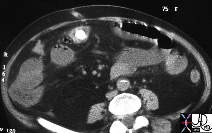 31118 31118 |
| 31118 hx 75 F small bowel fx dilated fluid filled fx calcification dx SBO gallstone ileus CTscan Courtesy Ashley Davidoff MD DB |
 31123 31123 |
| 31123 hx 75 F small bowel fx dilated gallbladder fx calcifcation fx air dx SBO gallstone ileus CTscan Courtesy Ashley Davidoff MD DB |
 Hyperplastic Cholecytoses Aschoff Rokitansky Sinus Hyperplastic Cholecytoses Aschoff Rokitansky Sinus |
| 19701 gallbladder + wall + fx calcification + cholecystoses + hyperplastic cholecystoses cholesterolosis imaging radiology CTscan Aschoff -Rokitansky sinuses |
Dx Hyperplastic Cholecystoses
Adenomyomatosis is an idiopathic, tumor-like abnormality of the gallbladder that is manifested by various hyperplastic changes such as overgrowth of the mucosa and thickening of the muscular wall. There is also development of intramural diverticulae, crypts, and sinus tracts within the gallbaldder wall called Rokitansky-Aschoff sinuses. These sinuses may be visible on ultrasonography by demonstration of ring-down artifacts that appear like “comet-tails”.
There is no malignant potential and the condition may involve the gallbladder in a focal, segmental, or diffuse form. Differentiation from gallbladder carcinoma may be difficult in some cases.
 19385 19385 |
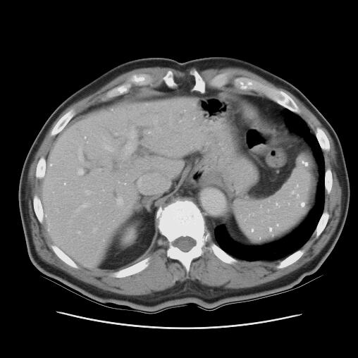
| 19385 liver + fx calcification + granulomatous disease + imaging radiology CTscan infection hot seat TB histoplasmosis code bile duct dilated |
 19394c01 19394c01 |
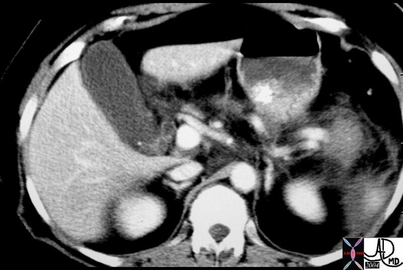
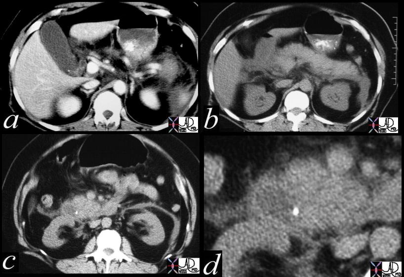
| 19394c01 code pancreas pancreatic head uncinate process fx mass fx central calcification dx serous cystadenoma code pancreatic duct fx dilated code fx calcifications dx granulomatous disease spleen code kidney fx small + imaging radiology CTscan code mass neoplasm benign tumor |
 32913 32913 |
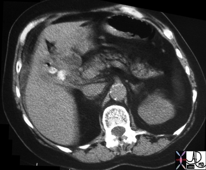
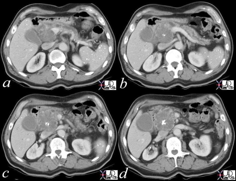
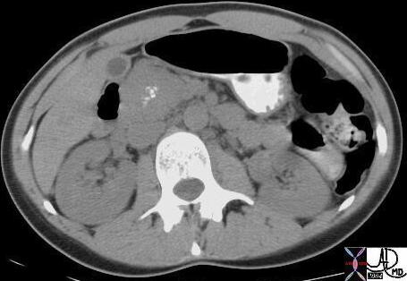
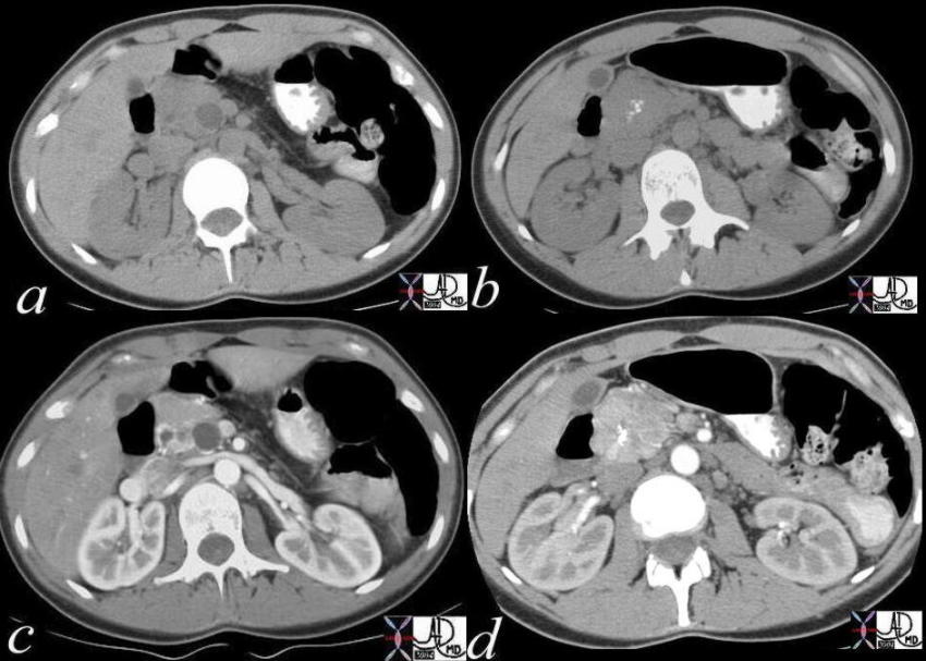
| 32913 Courtesy Ashley Davidoff MD abdomen calcification pancreas calcified body tail uncinate process chronic pancreatitis dystrophic calcification medical students inflammation imaging radiology KUB radiologists and detectives |
 32915b01 32915b01 |
| 32915b01 Courtesy Ashley Davidoff MD abdomen calcification pancreas calcified body tail uncinate process chronic pancreatitis dystrophic calcification medical students inflammation imaging radiology KUB radiologists and detectives |
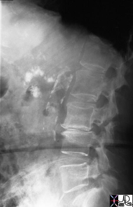
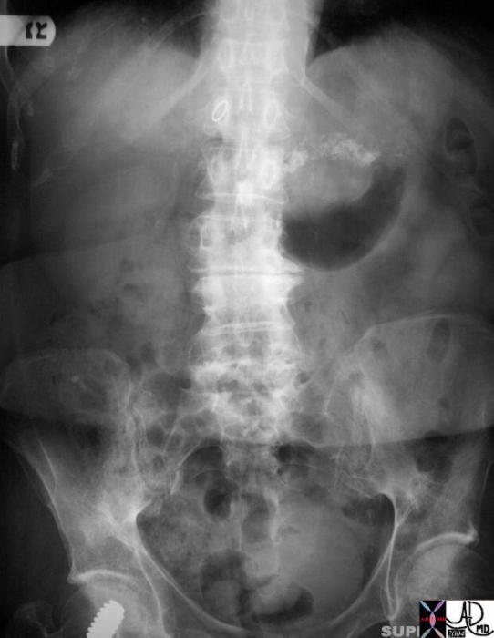
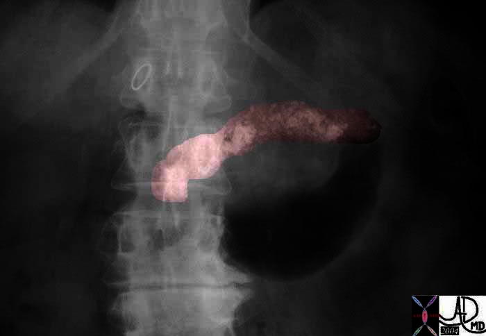
| 32916 |
| 32916 Courtesy Ashley Davidoff MD abdomen calcification pancreas calcified body tail uncinate process chronic pancreatitis dystrophic calcification medical students inflammation imaging radiology lateral abdomen aorta radiologists and detectives |
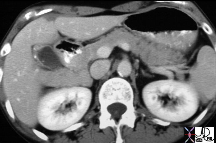
| 26076 |
| 26076 Courtesy Ashley Davidoff MD code pancreas code bile duct + fx filling defects + fx calcifications + dx cholelithiasis + dx gallstone pancreatitis + imaging radiology CTscan C+ |
| 26079c01 |
| 26079c01 Courtesy Ashley Davidoff MD code pancreas + fx indurated + dx gallstone pancreatitis + imaging radiology CTscan C+ code bile duct + fx filling defects + fx calcifications + dx cholelithiasis + dx gallstone pancreatitis + imaging radiology |
| 20889 |
| 20889 code spleen + hilum + capsule + fx calcification + metastatic primary ovary + carcinoma + imaging radiology CTscan C- liver parenchyma capsule + fx calcification + nodule metastatic primary ovary ligamentum venosum fx calcium calcification dx metastatic primary ovary neoplasm malignant metastasis metastases cancer tumor skin third spacing pleural effusion |
| 16868 |
| 16868 diaphragm liver porta hepatis gastrohepatic ligament Glissons capsule + fx calcification + primary ovary + metastasis + imaging radiology CTscan neoplasm malignant carcinoma tumor cancer |
| 16869 |
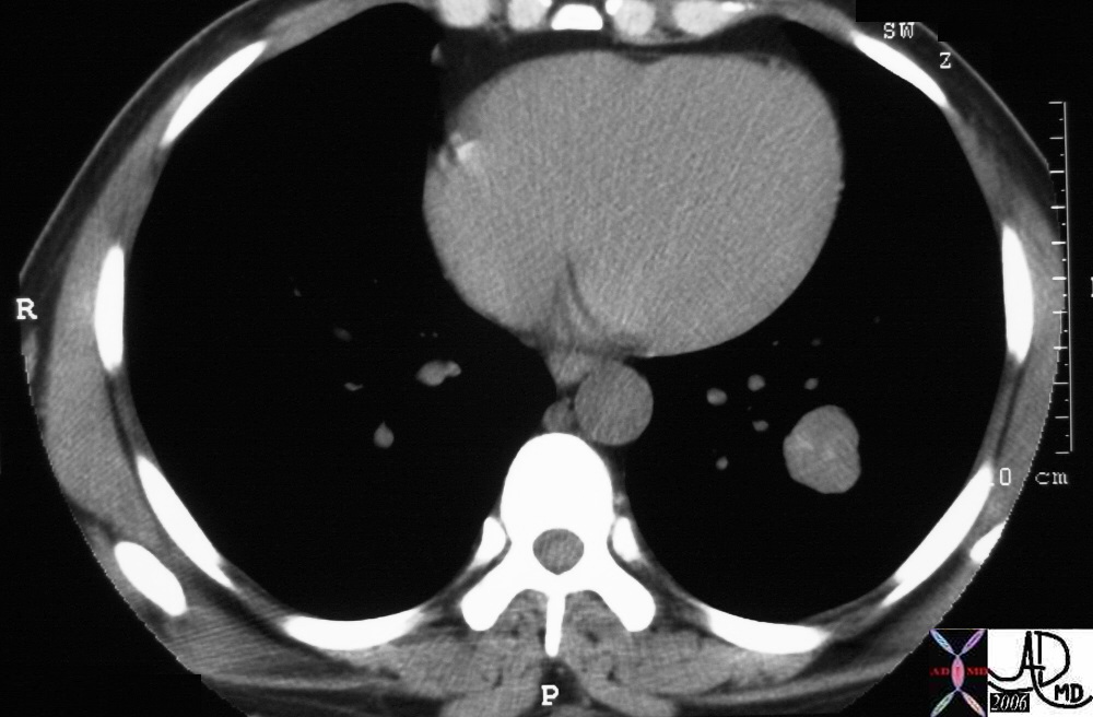
| 16869 Punctate calcifications on the liver capsule pleura and diaphragm are noted on this chest CT. The patient has metastatic ovarian carcinoma to the pleura and pericardium with calcification. Courtesy Ashley Davidoff MD. code diaphragm liver porta hepatis gastrohepatic ligament Glissons capsule + fx calcification + primary ovary + metastasis + imaging radiology CTscan neoplasm malignant carcinoma tumor cancer spleen splenic |
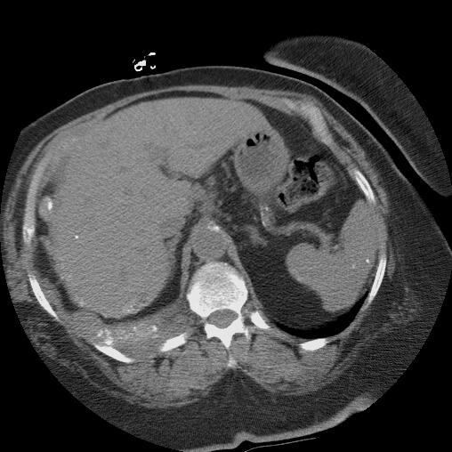
| 22357.800 |
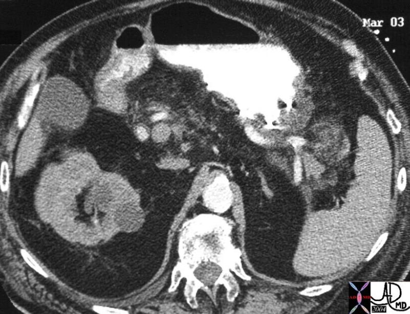
| 22357.800 51 M hepatic bare area liver abdomen ascites fx mass fx stippled calcification calcified fx psammomatous psamomatous calcifications dx metastattic mucinous adenocarcinoma of the rectum CTscan soft tissue windows Davidoff MD 22359 22357.800 |
| 22359 |
| 22359 51 M hepatic bare area liver abdomen ascites fx mass fx stippled calcification calcified fx psammomatous psamomatous calcifications dx metastattic mucinous adenocarcinoma of the rectum CTscan soft tissue windows Davidoff MD 22359 22357.800 |
| 18126 |
| 18126 liver + fx mass + fx calcification + bile duct + cholangiocarcinoma + imaging radiology CTscan splenic early heterogeneous contrast enhancement |
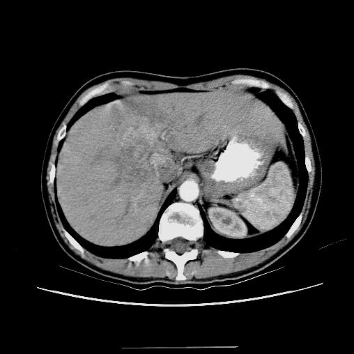
Gallbladder
| 16225 |
| 16225 Courtesy Ashley DAvidoff MD code gallbladder + infundibulum fx calcification dx cholelithiasis code cystic duct fx dilated fx enhancingwall dx obstruction code abdomen retroperitoneum Gerota’s fascia fx indurated dx gallstone pancreatitis + imaging radiology CTscan |
| 16225c |
| 16225c Courtesy Ashley Davidoff MD code gallbladder + infundibulum fx calcification dx cholelithiasis code cystic duct fx dilated fx enhancingwall dx obstruction code abdomen retroperitoneum Gerota’s fascia fx indurated code pancreas pancreatic uncinate process fx indurated bile duct fx dilated fx enhancing wall fx calcification stone dx choledocholithiasis dx gallstone pancreatitis + imaging radiology CTscan |
| 18378 |
| 18378 Courtesy Ashley Davidoff MD code pancreas + fx mass + fx calcification + serous cystadenoma von Hippel Lindau + imaging radiology CTscan C- |
|
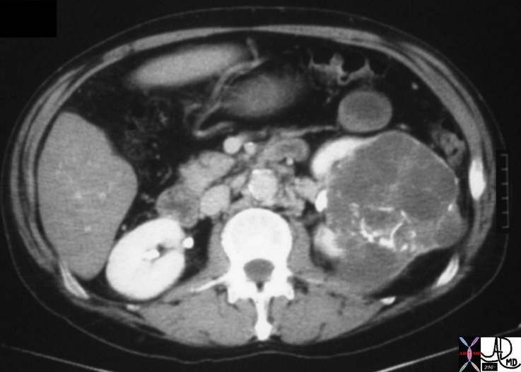
Cystic Renal Celll Carcinoma |
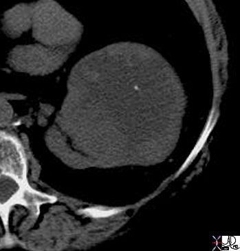
| 05730.800 kidney renal mass fx coarse calcifications curvilinear linear calcified space occupying displacement cystic enhancing septations dx cystic renal cell carcinoma RCC CTscan Davidoff MD Bosniak grade 4 05730.800 05730b.800 05729b |
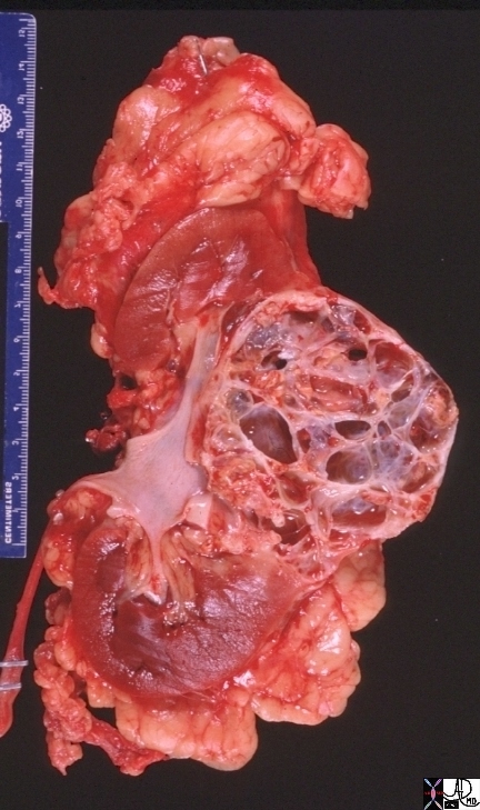
Cystic Renal Cell Carcinoma |
| 05729b kidney renal mass space occupying displacement cystic dx cystic renal cell carcinoma RCC grosspathology CTscan Davidoff MD Bosniak grade 4 05730.800 05730b.800 05729b |
Nephrolithiasis and Milk of Calcium Urine |
| 16045 kidney renal nephrolithiasis fx calcifications calcified fluid fluid level dx milk of calcium urine CTscan Davidoff MD 16046b01 16046 |
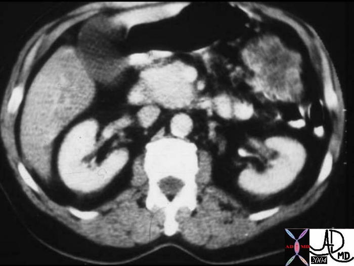
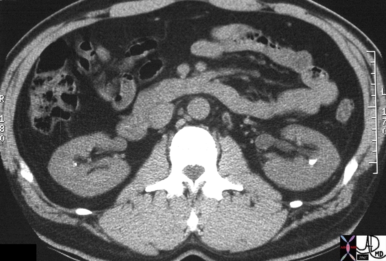
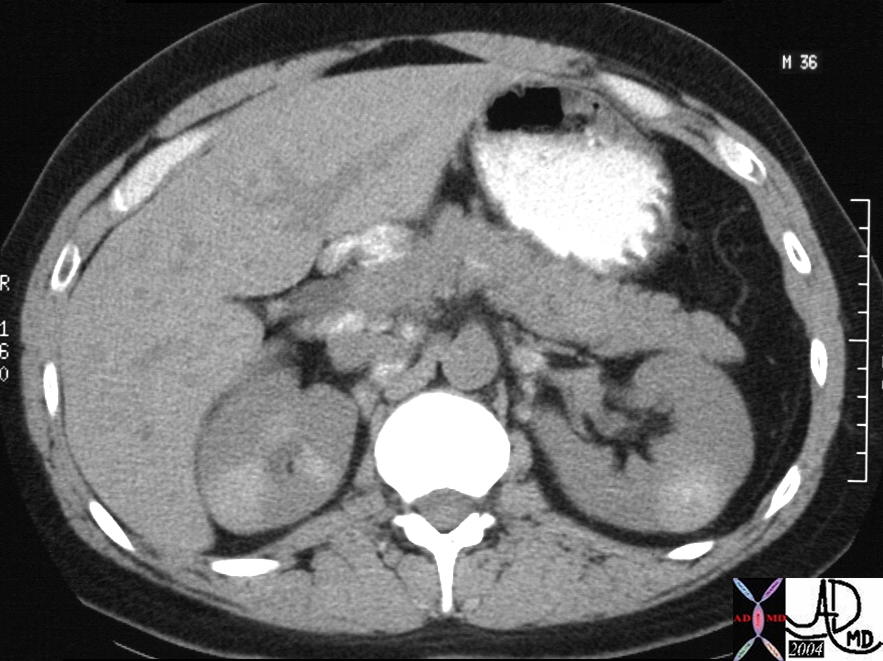
| 16808 |
| 16808 kidney fx renal mass fx punctate calcification RCC renal cell carcinoma malignant cancer pancreas head and uncinate process aging fatty atrophy of dorsal portion CT scan |
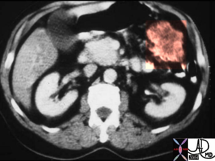
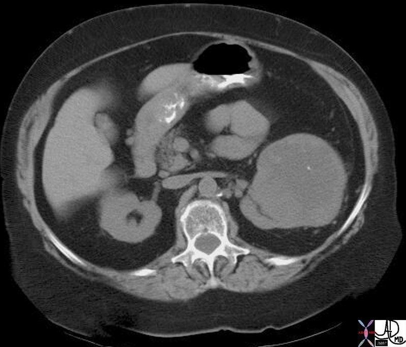
| 16808b01 |
| 16808b01 kidney fx renal mass fx punctate calcification RCC renal cell carcinoma malignant cancer pancreas head and uncinate process aging fatty atrophy of dorsal portion CT scan |
| 26477b02 |
| 26477b02 Courtesy Ashley Davidoff MD pancreas anatomy lymph node lymphatic drainage fx calcified calcification dx sarcoidosis kidney mass calcification calcified imaging radiology CTscan |
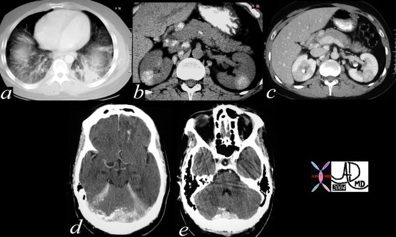
| 40406c |
| 40406c Courtesy Ashley Davidoff MD code lung fx interstium fx thickening dx interstitial lung disease ILD code pancreas abdomen lymph nodes fx calcified code brain meninges fx calcifications code dx sarcoidosis imaging radiology CT scan code inflammation immune |
Bladder
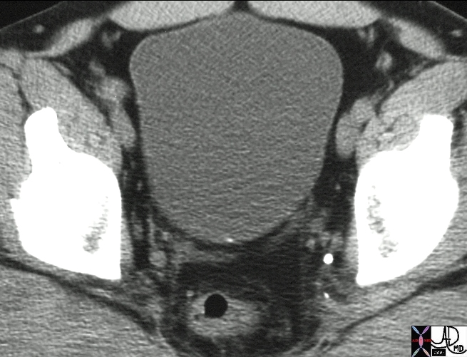
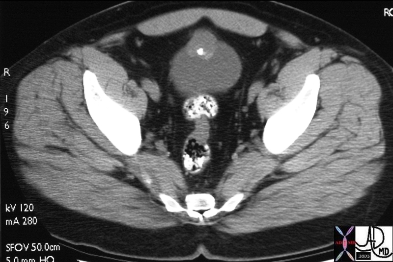
Is the stone in the Bladder or Ureter? Use of Gravity |
| 16046 urinary bladder fx calcification calcified lying posterior and midline dx recently passed stone CTscan Davidoff MD 16046 16046b01 |
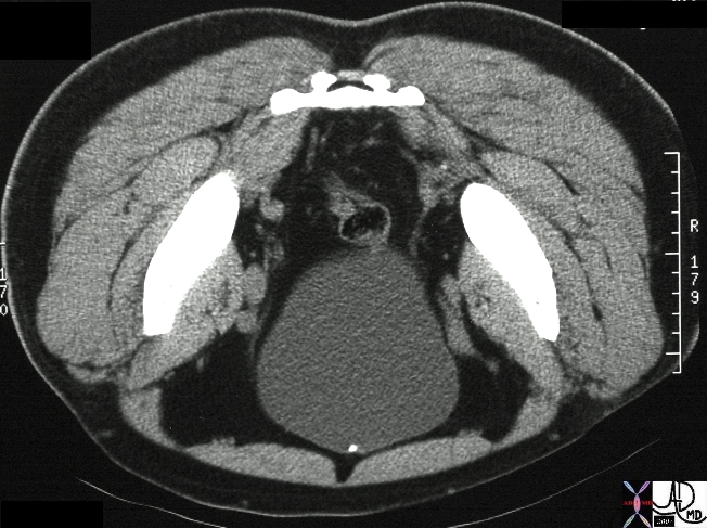
| 17108 |
| 17108 urinary bladder dome anterior wall calcification dx urachal remnant calcification CTscan Davidoff MD |
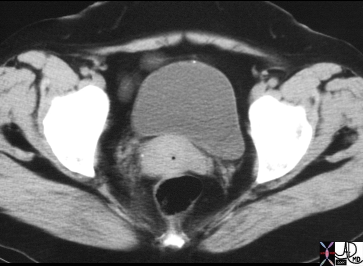
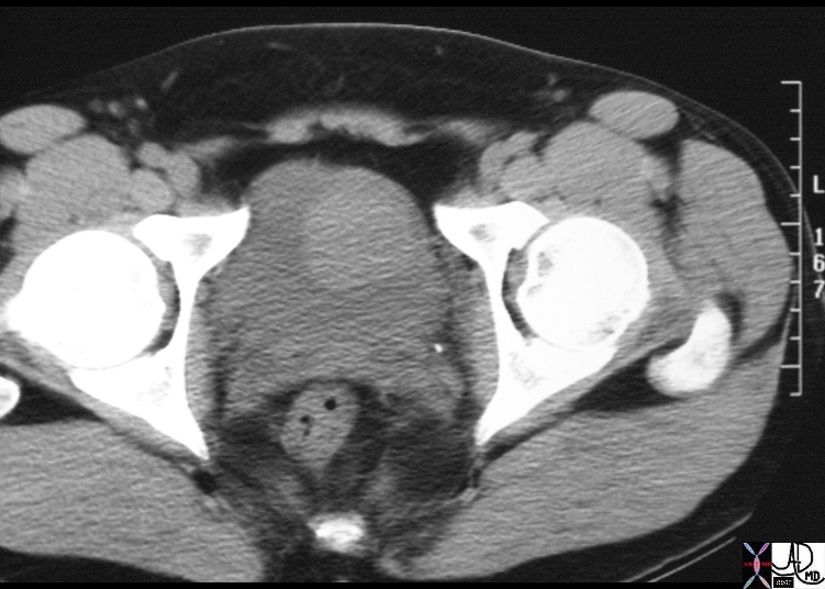
| 17102 |
| 17102 44F with hematuria fx anterior bladder mass fx calcification fx mass dx urachal adenocarcinoma imaging CTscan Courtesy Ashley Davidoff MD |
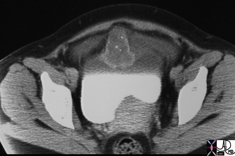
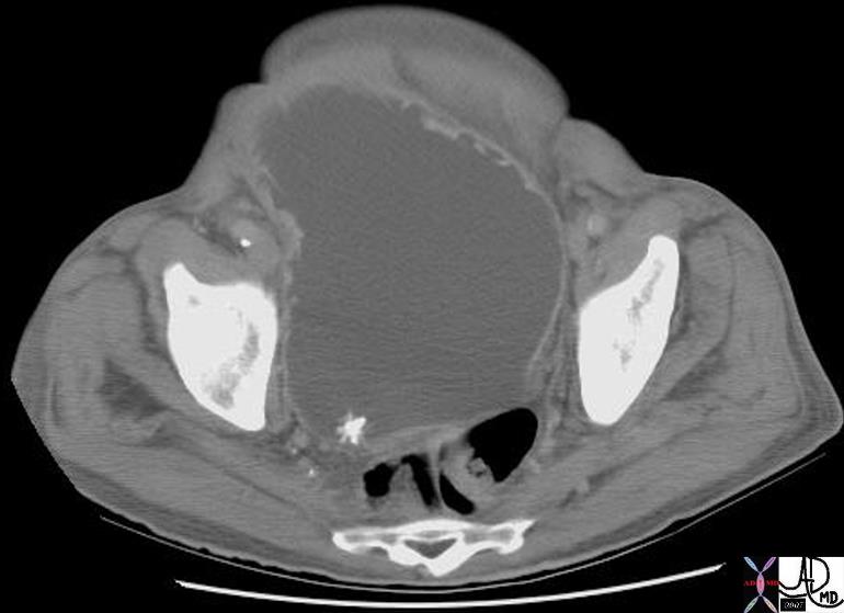
| 20068 |
| 20068 44M with hematuria fx anterior bladder mass fx calcification dx urachal adenocarcinoma imaging CTscan Courtesy Ashley Davidoff MD |
| 16568 |
| 16568 urinary bladder nodule fx relatively hypodense fx contrast in bladder dx bladder stone calculus CTscan Davidoff MD |
| 16758 |
| 16758 urinary bladder nodule fx relatively hypodense fx contrast in bladder dx bladder stone calculus CTscan Davidoff MD |
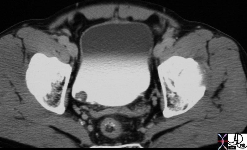
| 17103 |
| 17103 26M syncope on urination urinary bladder mass dense phlebolith dx phaeochromocytoma CTscan Davidoff MD 17106 17103 17104 17105 17106 17107 |
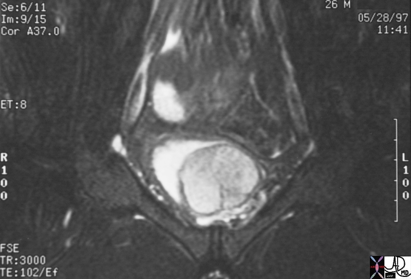
| 17106 |
| 17106 26M syncope on urination urinary bladder mass dense phlebolith dx phaeochromocytoma MRI T2 Davidoff MD 17106 17103 17104 17105 17106 17107 |
RES
| 20730b |
| 20730b spleen + splenic capsule calcified +calcification imaging radiology CTscan |
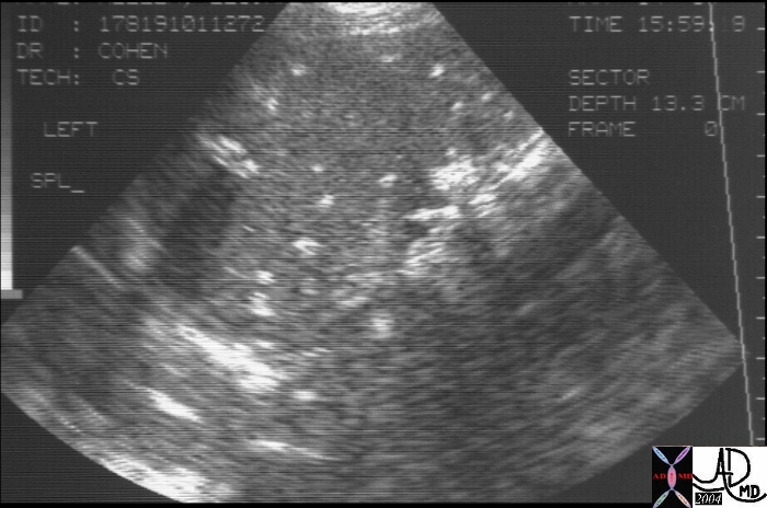
| 20996b |
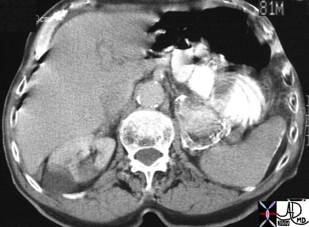
| 5 year old male 20996b spleen + fx calcifications multiple nodules + dx infarcts + imaging radiology USscan surgical proof |
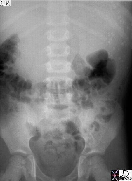
| 20995 |
| 20995 spleen + fx calcifications multiple calcified nodules + dx multiple splenic infarcts + plainfilm imaging radiology KUB X-ray |
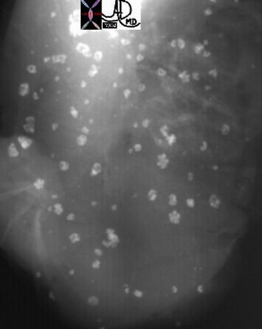
| 20997 |
| 5 year old male 20997 spleen + fx calcifications multiple + dx calcified splenic infarctions infarct + plainfilm surgical grosspathology |
| 20919a01 |
| 20919a01 spleen + dense thorotrast + celiac axis lymph nodes hyperdense calcific density plainfilm KUB X-ray abdomen infiltrative disorder RES reticuloendothelial system |
| 20919a03 |
| 20919a03 spleen + fx dense hyperdense fx nodule dx Thorotrast + imaging radiology CTscan C- non contrast liver capsule parenchyma calcific density calcification calcified gastrohepatic ligament lymph node fx hyperdense calcified infiltrative disease retciculoendothelial system RES rule out angiosarcoma |
| 20921 |
| 20921 spleen + fx dense hyperdense parenchyma dx Thorotrast + imaging radiology CTscan C- non contrast liver dense hyperdense parenchyma calcific density calcification calcified infiltrative disease retciculoendothelial system RES |
| 20924 |
| 20924 gallbladder fossa GBF + fx calcification + Thorotrast + imaging radiology CTscan liver dense hyperdense parenchyma calcific density calcification calcified infiltrative disease retciculoendothelial system RES |
| 20914 |
| 20914 hx 75F lymph node fx large calcified metastaic dx carcinoid tumor imaging radiology CTscan C- Courtesy Ashley Davidoff MD DB |
- Rarely, large osteoma in the frontal or ethmoid region may displace globe forward and cause proptosis
- Obstruction of a sinus ostium may lead to infection or formation of a mucocele
- Very rarely, an osteoma may erode through the dura leading to cerebrospinal fluid rhinorrhea or intracranial infection
Multiple osteoma of the mandible and maxilla, along with the frontal, sphenoid and ethmoid sinuses, rarely the long bones or phalanges
Cutaneous and soft tissue tumors
Association between colonic polyps with a predilection to malignant degeneration
Other Images
References
Gardners Syndrome – eMEdicine
Cardiovascular System
 15401 15401 |
| This CTscan with contrast through the heart shows heavy calcification of the LAD as well as the circumflex coronary artery as well as calcification of the aortic annulus. Courtesy Ashley Davidoff MD. 15401 code cardiac heart artery coronary calcification calcium calcified CAD atherosclerosis aortic sclerosis annulus imaging radiology CTscan AO aorta |
| Pericardial Calcification |
| 15527 heart pericardium fx calcification serous pericardium fibrous pericardium dx calcific pericarditis probable viral in origin imaging radiology CTscan Courtesy Ashley Davidoff MD |
| Pericardial Calcification |
| 29157 hx 55M with cough fx heart pericardium epicardium visceral pericardium parietal pericardium fx calcification dx calcific pericarditis probable viral virus imaging radiology plain film CXR Courtesy Ashley Davidoff MD |
 Amyloidosis Amyloidosis |
| 15528 heart + pericardium + fx calcification + amyloid + imaging radiology CTscan infiltrative amyloidosis |
 Amyloidosis Amyloidosis |
| abdomen + pericardium + fx calcification + amyloid + imaging radiology CTscan infiltrative amyloidosis |
 LV Apical Aneurysm LV Apical Aneurysm |
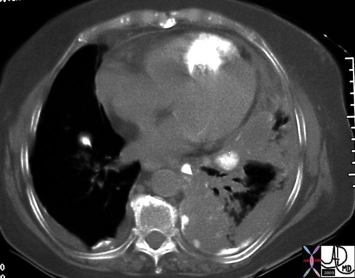
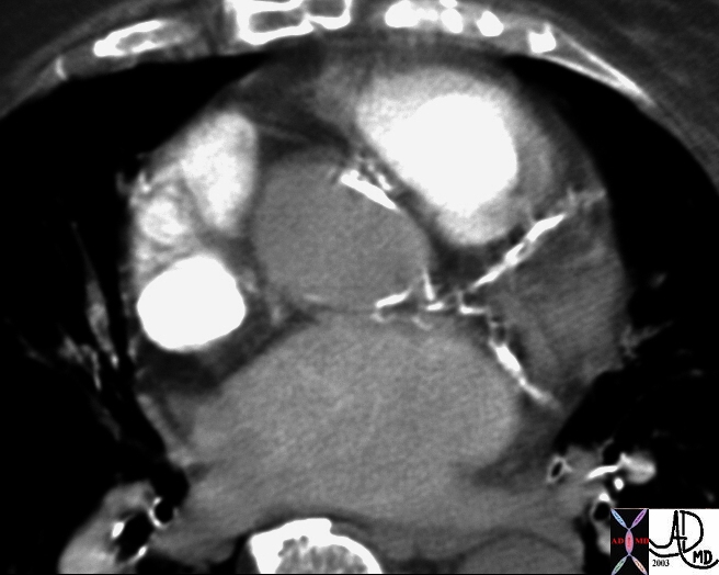
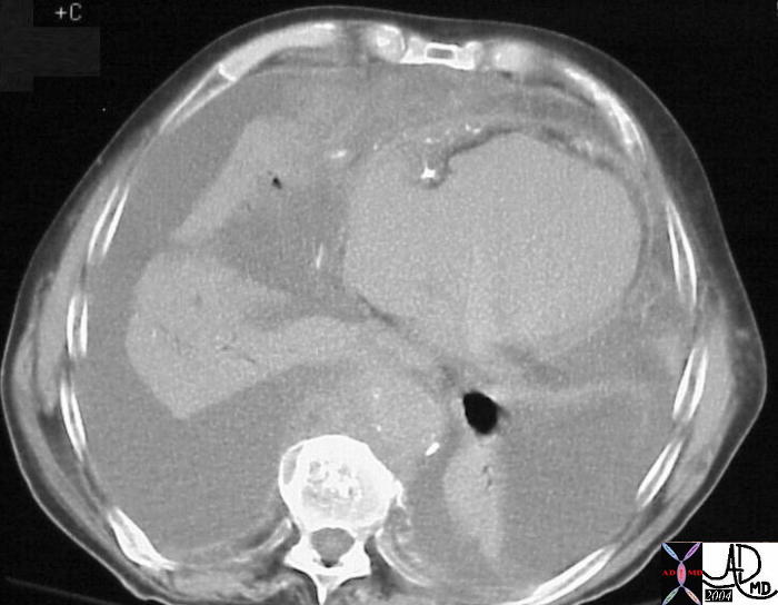
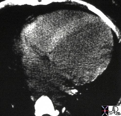
| This cross sectional CT image of the heart shows calcified apex of the left ventricle associated with thrombus, characteristic of an LV aneurysm. The cause is almost certainly secondary to coronary artery disease and ischemic heart disease with secondary myocardial infarction.
Courtesy Ashley Davidoff MD. 30472 code heart LV apex IHD CAD aneurysm calcification calcified MI cardiac imaging radiology CTscan |
| LV Apical Aneurysm Post Infarction |
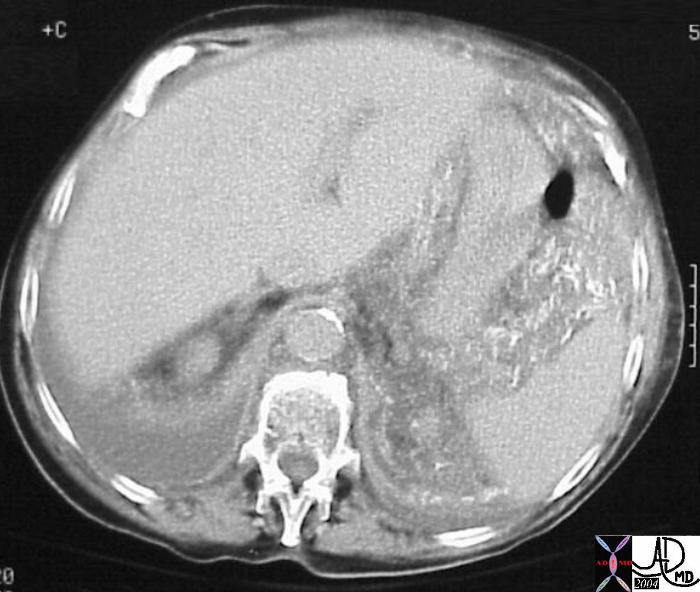
| This single cross sectional view of the heart through the LV shows apical myocardial dystrophic calcification. The pericardium can be seen as a fine soft tissue density between the two layers of fat – the epicardial fat (inner layer) and the pericardial fat. This finding is diagnostic of a previous MI and may represent dystrophic calcification in the myocardium or within thrombus in an aneurysm of the apex. Courtesy Ashley Davidoff MD. 29600 code CVS cardiac heart MI apex calcification calcified IHD myocardium cardiac imaging radiology CTscan |
 29601 29601 |
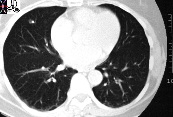
| This single axial CT image shows a faint lucurvilinear lucency in the apical myocardium. This lucency is thought to be a fatty deposition in infarcted myocardial tissue. The calcification in the annulus is premature and unusual for this 56 year old male patient. Note the small bilateral pleural effusions. Courtesy Ashley Davidoff MD. 29601 code CVS cardiac heart MAC apical fat MI cardiac imaging radiology CTscan |
Papillary Muscle |
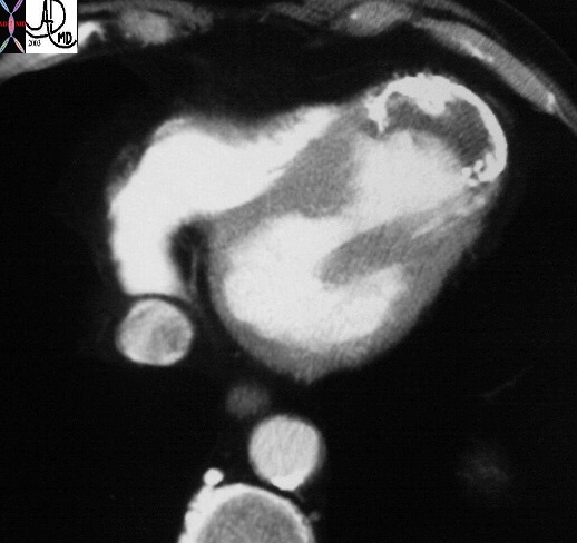
| The chest CT is taken through the heart with the contrast phase in the right sided structures. A focal calcification in the cavity of the left ventricle (LV) is seemingly related to a focal hypodensity of the paillary muscle and may represent a calcific nodule on the chotdae or papillary muscle tip. Courtesy Ashley Davidoff MD 26320 code LV papillary muscle calcification chordae tendinae cardiac imaging radiology CTscan |
| 24672 |
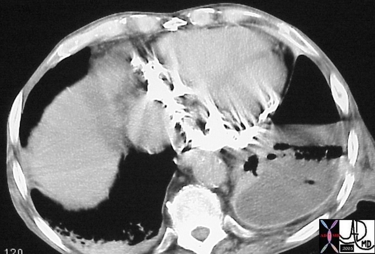
| This is a CT of the chest at the level of the heart filmed on narrow windows showing a dense ventricular septum. The autopsy specimen showed calcific myocarditis. Courtesy Ashley Davidoff MD 24672 code heart septum dense calcific calcified calcification myocarditis inflammation cardiac imaging radiology CTscan |
| 15529 |
| 15529 abdomen + gastrolienal ligament +spleen stomach fx calcification + amyloid + imaging radiology CTscan amyloidosis infiltrative |
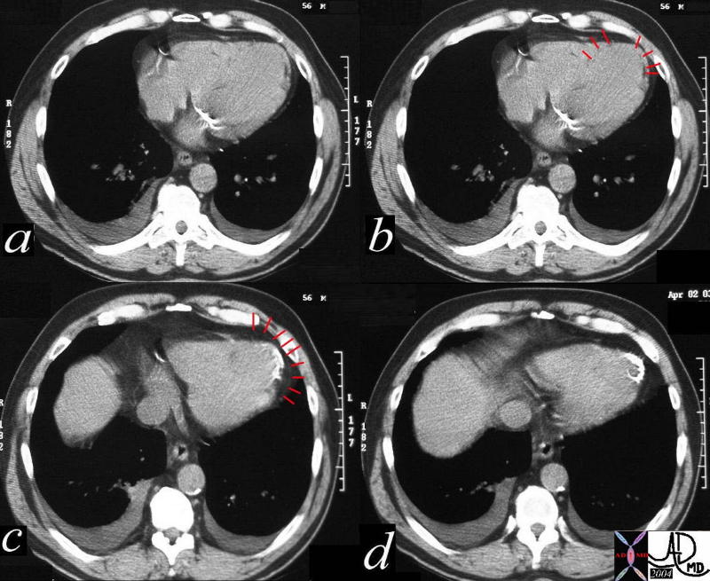
19426c |
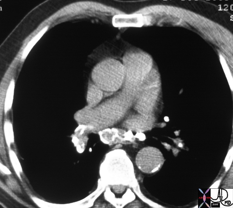
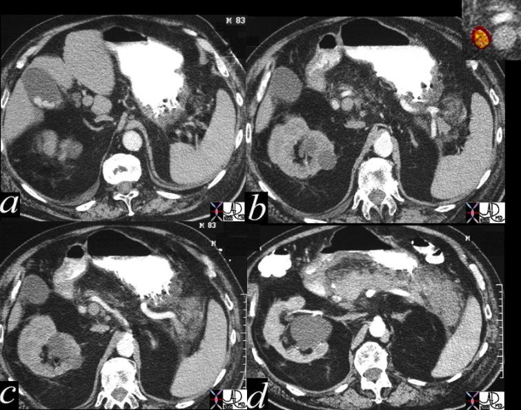
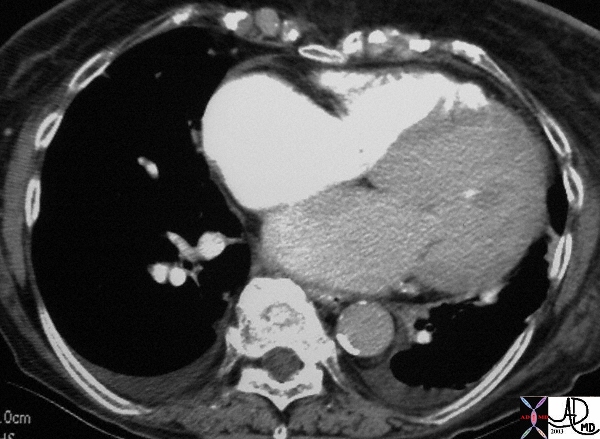
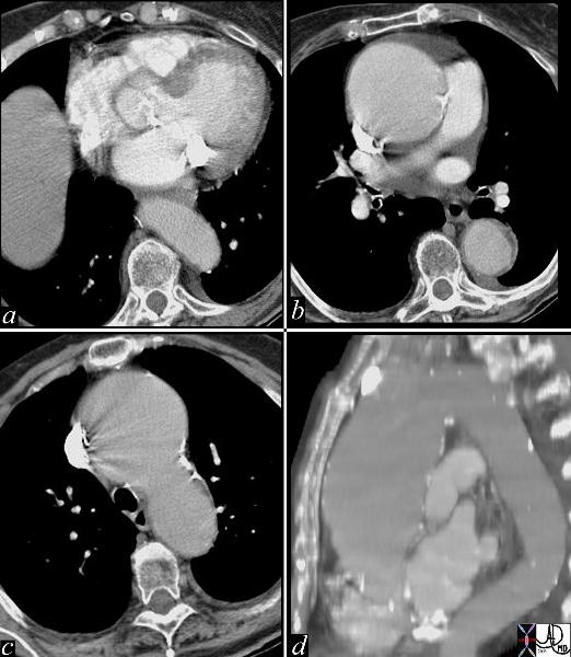
| This series of images are from a CTscan showing an ascending aortic thoracic aneurysm. There is evidence of heavy calcification of the aortic valve (aortic sclerosis), an aneurysm confined to the ascending aorta (b,c,d), and tortuosity of the descending aorta (d). The cause for the aneurysm is probably a combination of systemic hypertension, aortic stenosis and atherosclerotic degeneration of the wall. Courtesy Ashley Davidoff MD 19426c code CVS thorax aorta ascending aneurysm MAC aortic sclerosis CTscan |

22734 cW02 |
| This angiogram of the abdominal aorta shows a widened infrarenal aorta. At first glance the lumen of the aorta appears normal, but a faint curvilinar calcification of the true wall can be seen to the patients left in the first image. The second image (b) reveals the true size of the aneurysm. Courtesy Ashley Davidoff MD 22734 cW02 codeCVS aorta artery abdomen aneurysm AAA |
33289 |
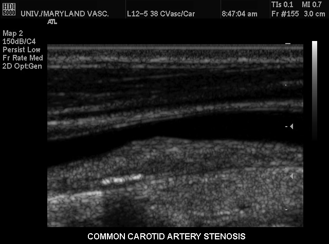
| This conventional gray scale US of the neck shows a common carotid artery heaped up plaque on the far wall causing stenosis of moderate degree. Note also the linear area of strong echogenicity on the far wall associated with shadowing consistent with a calcification in the wall. These findings are characteristic of atherosclerotic plaque. Courtesy Philips Medical Systems 33289 |
Gardner’s syndrome |