Copyright 2007
Principles
Infection
Cysticercosis |
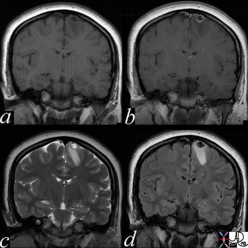
| 71575c02 24 female presents with seizures brain cortex vertex parafalcine falx fx nodule 9mm ring thinly enhancing lesion left frontal lobe with tiny mural nodule which enhances as well rim T2 hypointense ? calcification nodule T2 hyperintense white matter edema edema right frontal lobe a = T1 pre gadolinium precontrast b= T1 – post contrast post gadolinium c= T2 weighted image d = FLAIR dx cysticercosis infection Courtesy Davidoff MD |
CNS
|
Cysticercosis |
| 71575c02 24 female presents with seizures brain cortex vertex parafalcine falx fx nodule 9mm ring thinly enhancing lesion left frontal lobe with tiny mural nodule which enhances as well rim T2 hypointense ? calcification nodule T2 hyperintense white matter edema edema right frontal lobe a = T1 pre gadolinium precontrast b= T1 – post contrast post gadolinium c= T2 weighted image d = FLAIR dx cysticercosis infection Courtesy Davidoff MD |
CVS

Sarcoidosis of the Heart |
| 49740c01 34 male with sarcoidosis presents with dizzineess and complete heart block cardiac heart LV sarcoidosis complete heart blosk early gadolinium delayed gadolinium enhancement of the LV wall left ventricle inflammation MRI T1 Davidoff MD |
RS
GIT
Infection
Hypervascular Lesion in the Spleen Blastomycosis |
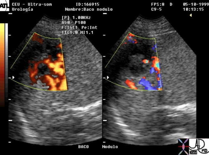
| 22381 spleen + fx echogenic splenic nodule hypervascular mass blastomycosis + imaging radiology USscan color doppler infection |
GUT
MSK
RES
Skin