copyright 2007

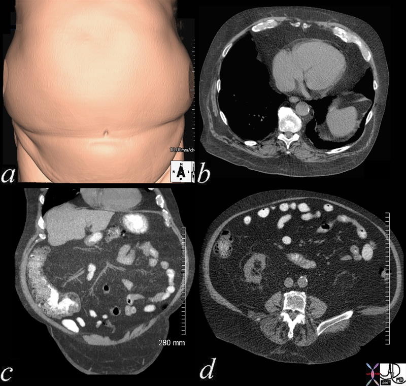
Round in Disorder |
| 49671c01 abdomen fat adipose tissue storage rotund belly umbilicus belly button surface rendring CTscan Davidoff MD |
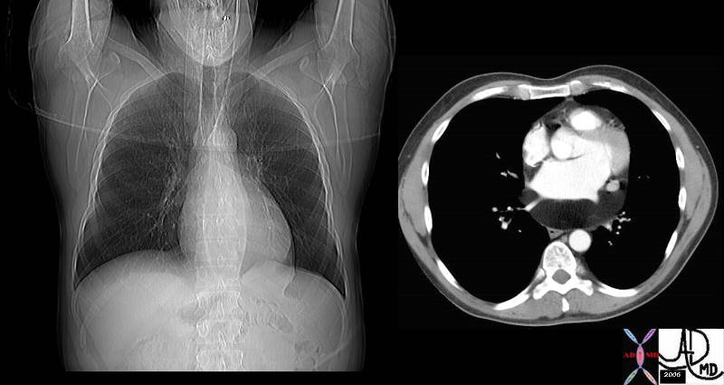
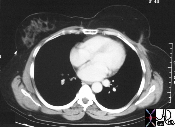
Cushing’s Syndrome – Iatrogenic |
| 71194c01 hx patient being treated on steroids abdomen pericardium mediastinum fat adipoase tissue centripetal centripedal distribution of adiposity pericardial fat mediastinal fat rotund shape size accumulation rounded dx Cushings syndrome iatrogenic CTscan #D volume rendering surface rendering Davidoff MD abdominal fat adiposity |

Morbid Obesity |
| 49715 woman adipose tissue obesity small on the inside large on the outside fat storage CTscan scout Courtesy Ashley Davidoff MD |

Body in Health and Disease |
| 49609c02 abdomen health disease normal obese morbid obesity order disorder Davidoff MD |
CVS
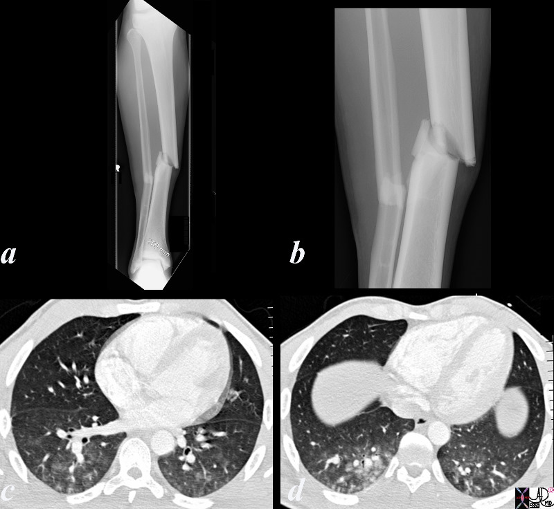
Lipoma of the Pericardium |
| 34800c01 cardiac heart pericardium fx lucent subcarinal angle altered dx lipoma of pericardium fat CTscan plain film scout Davidoff MD |
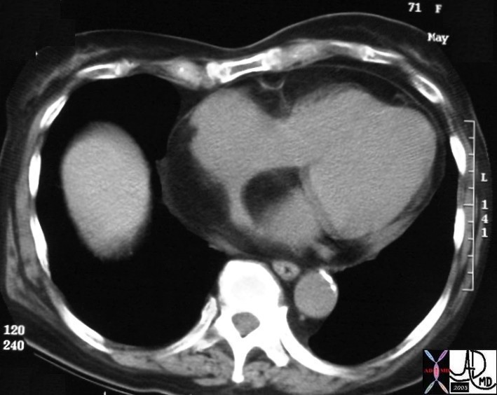
Lipoma of the Interatrial Septum |
| 28386 71F on steroids heart interatrial septum epicardial fat fx large fat accumulation dx interatrial lipoma imaging radiology CTscan Courtesy Ashley Davidoff MD |
Reconstructing the Shape of the Breast |
| 16127 Courtesy Ashley Davidoff MD code breast TRAM imaging radiology CTscan post mammoplasty |
RS

Fat in Disease – Lung and Heart |
| 49722c01.800 chest bronchi lungs atelectasis fat lipid lipoid pneumonia LV heart cardiac fat low density old chronic myocardial infarction CTscan Davidoff MD Courtesy Dr David Lee |
GIT
| Acute Cholecystitis Caused by Cholesterol Crystals |
| 77753c03.8s young female right upperquadrant tenderness RUQ gallbladder echogenic irregular fluid fluid layer conforming to the shape of the gallbladder wall thickened linear echoes floaters crystalline SG less than bile cholesterol crystals specific gravity forces space cholelithiasis stones small acute cholecystitis USscan ultrasound Courtesy Ashley Davidoff MD For Radiologists and Detectives |
Approach
Since the normal aging process of the pancreas results in fatty infiltration of the gland finding an echogenic or fatty pancreas on routine imaging is not uncommon. There is a range of normal from mild where the infiltrating fat creates a lobular appearance to the surface of the gland to severe infiltration where the pancreas is almost totally replaced by fat and the glandular tissue is visible as strands of tissue scattered throughout the gland. In the latter instance there is a surprising absence of functional significance. In patients with cystic fibrosis the pancreas is totally replaced and even strand of tissue are not seen. It is in these instances that functional changes may be seen. There are rare instance of lipomas in the gland which are easily recognized. Therefore fat in the pancreas in general does not present as a challenge in differential diagnosis. In the young patient with severe fatty change ask about cystic fibrosis, and in the patient with a lipoma, ask about associated syndromes. There are only a few isolated reports of lipomas in the literature.
Differential Diagnosis
aging process 99.9%
cystic fibrosis
lipoma
The transverse section through the pancreas shows an echogenic pancreas caused by infiltration of retroperitoneal fat into the age related involution of the pancreatic parenchyma. This is a normal and common finding, and usually has no clinical nor functional significance. 29495 Courtesy Ashley Davidoff MD
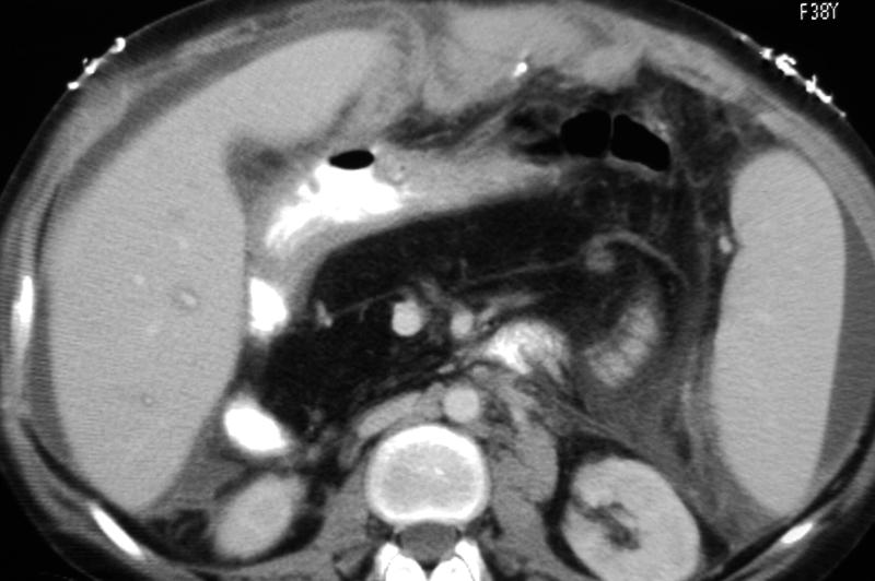
This CTscan through the tail of the pancreas shows involutional change of the tail of the pancreas characterized by the nodular appearance of the borders of the pancreas. This is a normal aging process whereby the involuting pancreatic parenchyma gets replaced by the retroperitoneal fat which has free access into the pancreas since there is no formal protective capsule surrounding the pancreas. 39807 Courtesy Ashley Davidoff MD
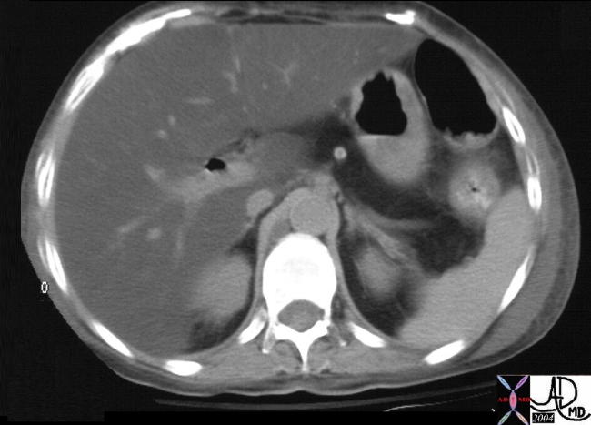
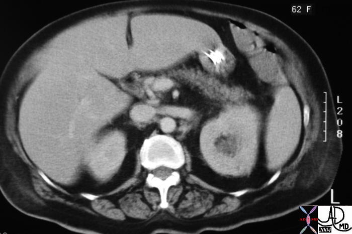
The CTscan through the body of the pancreas shows a calcification surrounded by a thin rim of soft tissue, just rightward of the poorly opacified SMV. The calcification represents a stone that is lodged in the distal CBD consistent with a diagnosis of choledocholithiasis. Incidental findings include the cysts in the right kidney. The pancreas has been almost totally replaced by fat which usually has no functional significance. 26076 Courtesy Ashley Davidoff MD
This CTscan through the pancreas shows a dominance of fat, with only connective tissue strands and blood vessels in this patient with cystic fibrosis. Note the complex ascites and thickening in the anterior pararenal space. 40625 Courtesy Ashley Davidoff MD
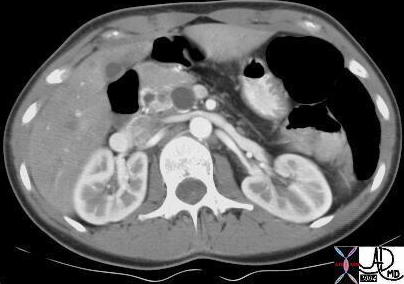
This CTscan through the liver and pancreas shows severe fatty change in the body and tail of the pancreas associated with severe steatosis of the liver. Air in the biliary system is of incidental note. This patient has cystic fibrosis. 22581 Courtesy Ashley Davidoff MD
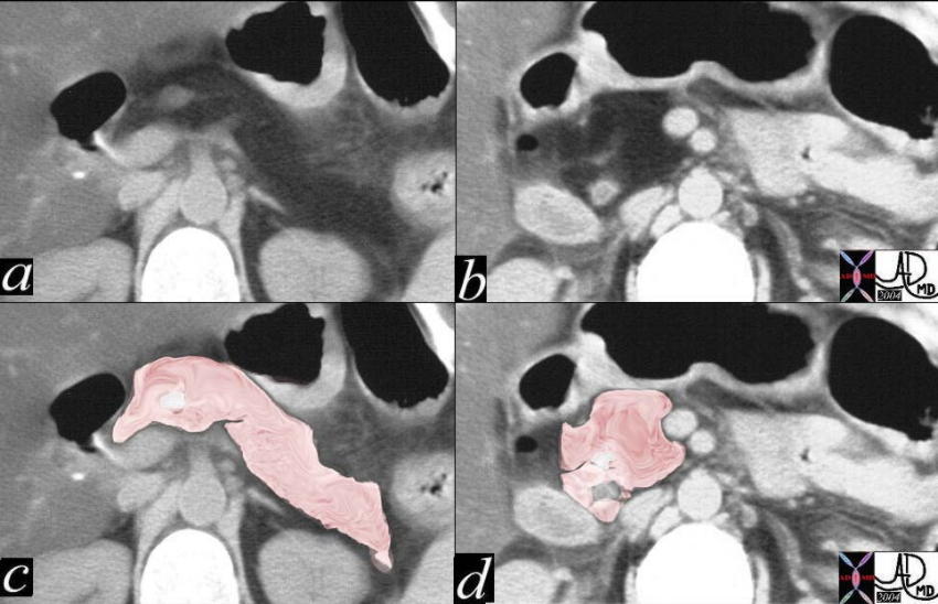
The following series reflects two levels of the case described above.
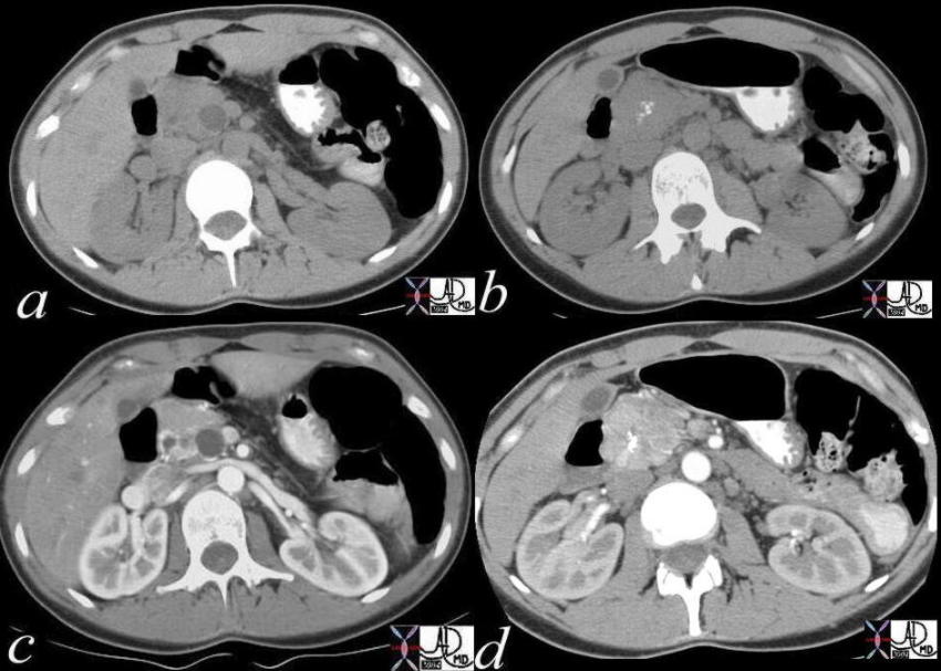
This CTscan through the liver and pancreas shows severe fatty change in the entire pancreas. Image a shows diffuse fatty change of the neck, body and tail, with image c showing a pink overlay of the pancreas. Image b shows a similar fatty infiltration in the head and uncinate process with image d representing the overlay of these structures. 22584c04 Courtesy Ashley Davidoff MD
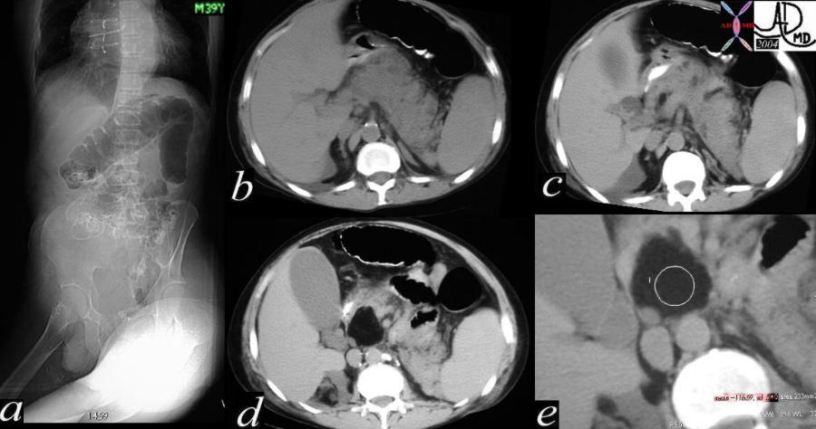
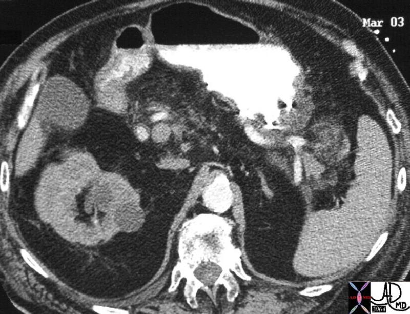
This CT scan shows an unusual pattern of fatty change of the pancreas in this patient with known von Hippel-Lindau syndrome. Noted is the severe fatty infiltration in the body and tail and total sparing of the neck and head. Associated findings include a variety of cysts in the uncinate process with both simple and complex characteristics. 18378b Courtesy Ashley Davidoff MD
The following series reflects two levels of the case described above both without and with contrast.

Mass with Fat, Calcium, Fluid Fluid Level – Dermoid of the Ovary |
| 24078 ovary fx mass fx fat fx calcification fx fluid-fluid level dx dermoid CTscan C- 24078 24077 Davidoff MD |
Musculoskeletal
|