Best Cases in Radiology
Copyright 2007

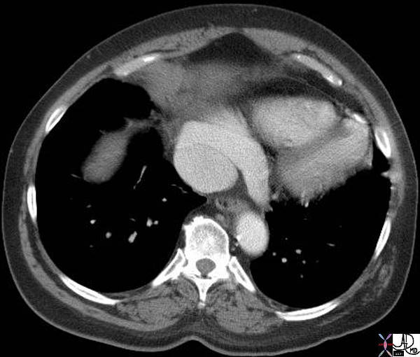
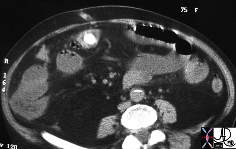
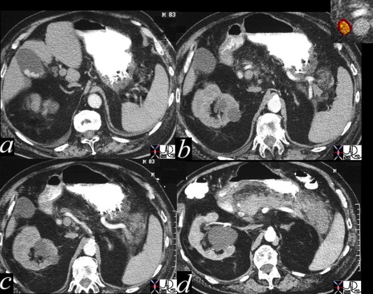
Massive Splenomengaly and….. |
| 00153 spleen enlarged splenomegaly portal hypertension vein SMV superior mesenteric vein fx calcification dx schistosomiasis schistosoma mansonii hepatic fibrosis cirrhosis imaging radiology CTscan |
Differential Diagnosis for Massive Splenomegaly
Infections
Malaria
Kala Azar
Schistosomiasis
Neoplastic Disease
Myelofibrosis Myeloid Metaolasia
Chronic Myeloid Leukemia
Large Splenic Cyst
Inherited Disorders
Thalassemia Major (Cooley’s Anemia)
Storage Disease – Gaucher’s and Niemann-Pick
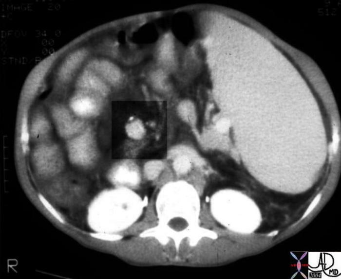
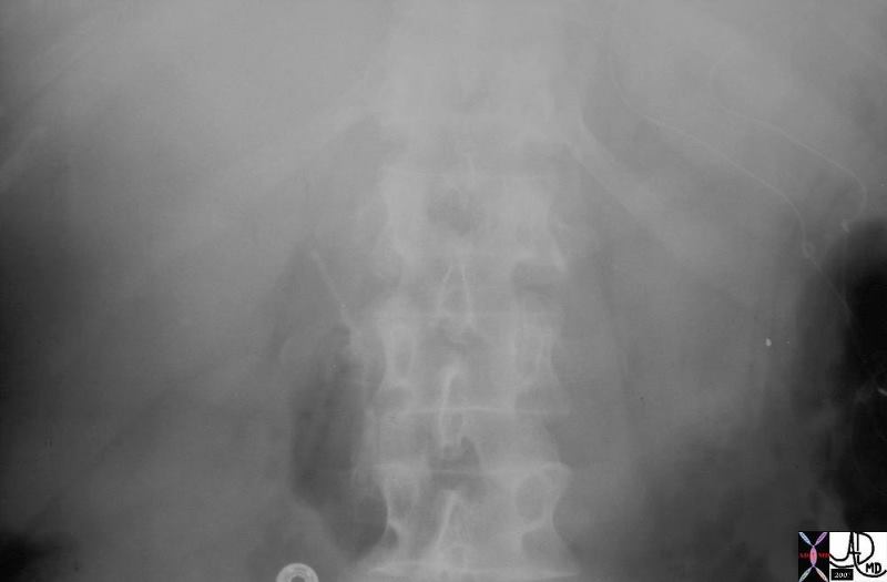
| Massive Splenomengaly and calcification in the Portal Vein |
| 00153 spleen enlarged splenomegaly portal hypertension vein SMV superior mesenteric vein fx calcification dx schistosomiasis schistosoma mansonii hepatic fibrosis cirrhosis imaging radiology CTscan |
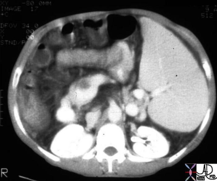
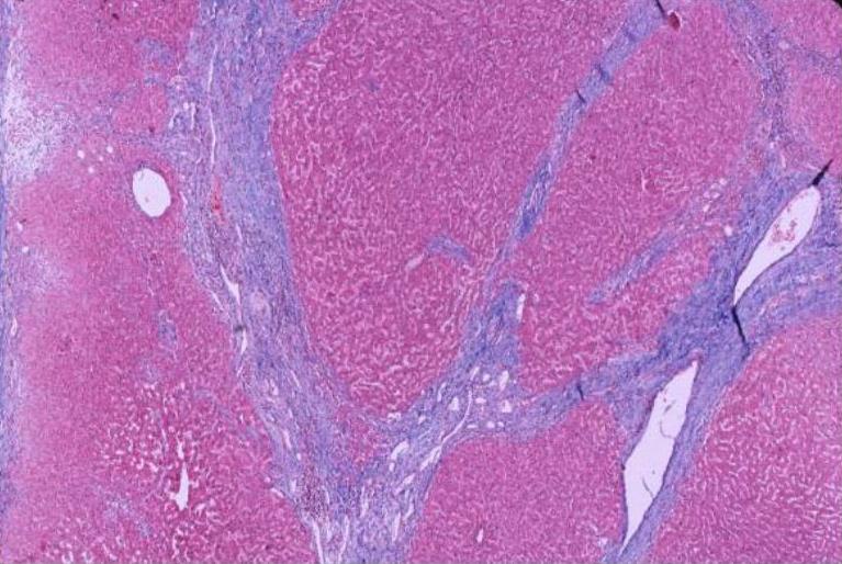
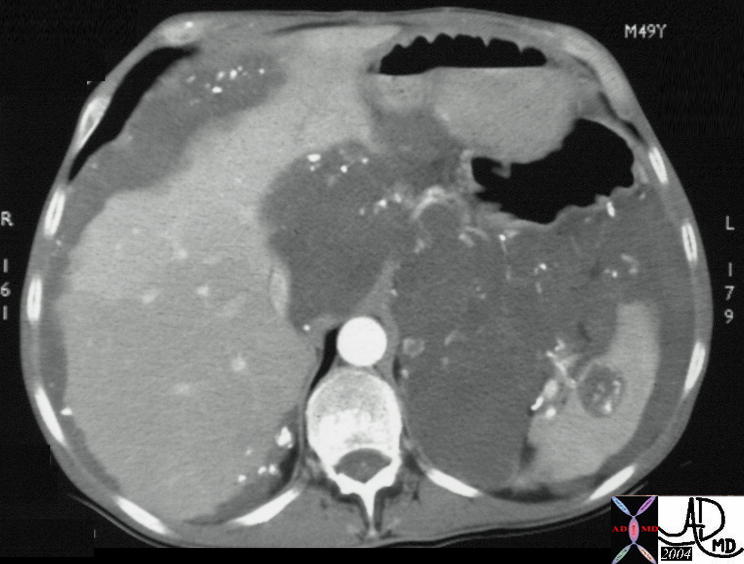
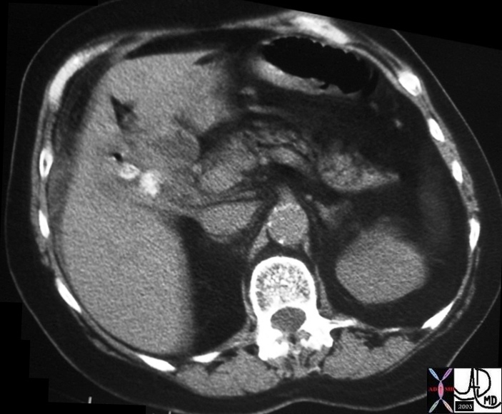
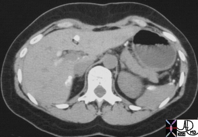
Liver – Bands of Fibrosis |
| 00153a01 liver + deformity + fx linear hypodense bands of fibrosis dx schistosomiasis + schistosoma mansonii imaging radiology CTscan infection cirrhosis portal hypertension |
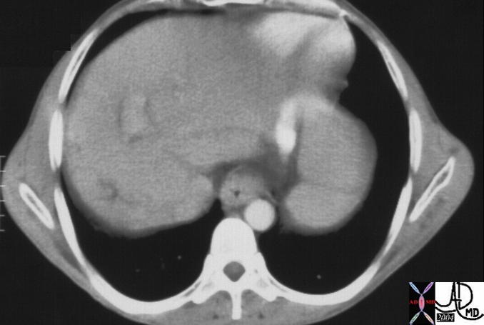
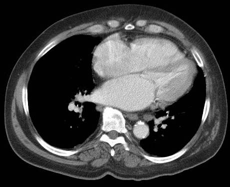
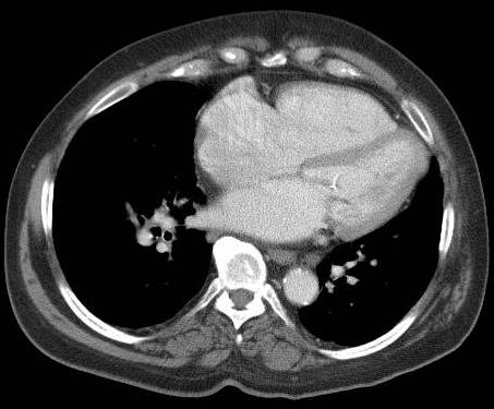
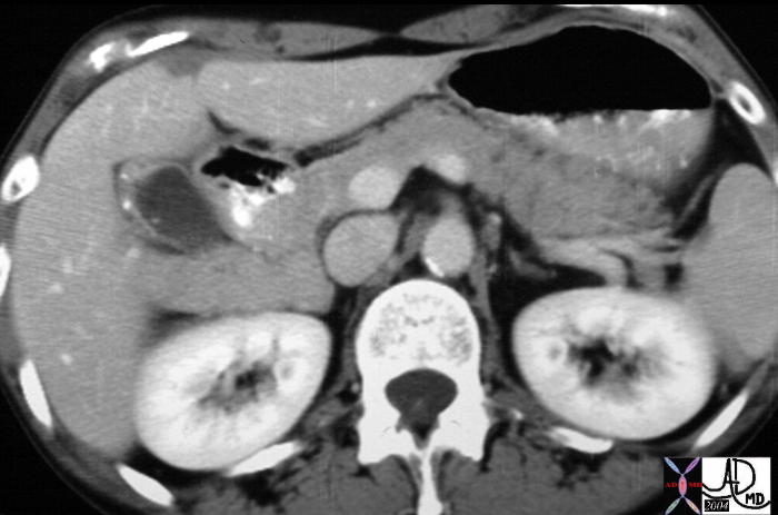
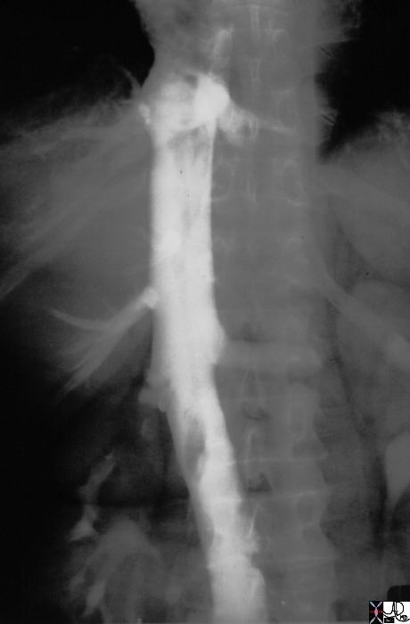
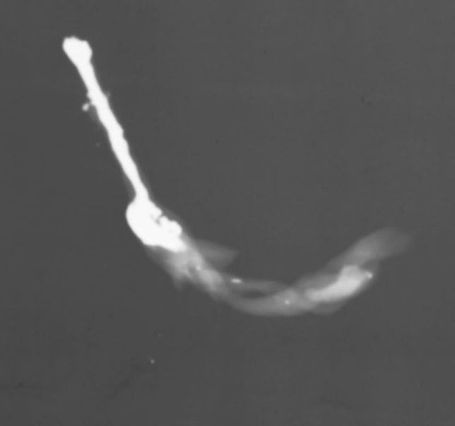
More Thrombus |
| 00154 spleen enlarged splenomegaly portal hypertension vein SMV superior mesenteric vein thrombosis fx dx schistosomiasis schistosoma mansonii hepatic fibrosis cirrhosis imaging radiology CTscan |
|
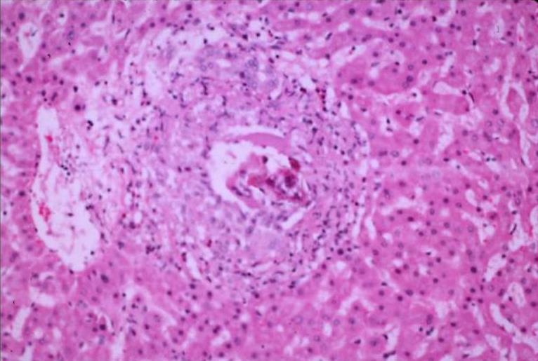
Granuloma in the Portal Vein with the Egg in the Center |
| 02273 02275 hx young man from Puerto Rico liver schistosomiasis schistosoma hematobium egg histopathology Courtesy Barbara Banner MD |
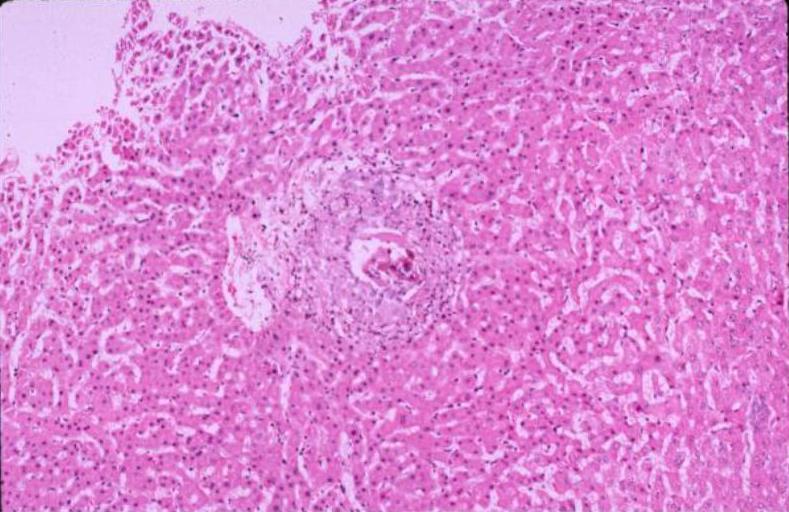
 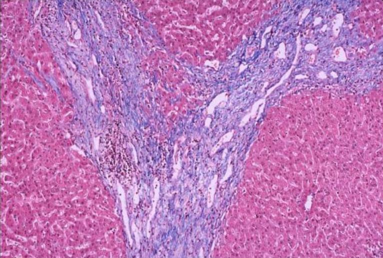
Bands of Fibrosis |
|
02270 02272 hx young man from Puerto Rico liver schistosomiasis hepatic cirrhosis histopathology Courtesy Barbara Banner MD portal fibrosis stands out , termed as “pipestem” fibrosis.
|

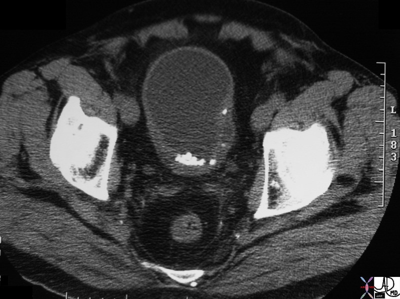
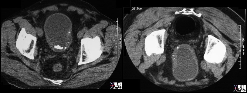
33289
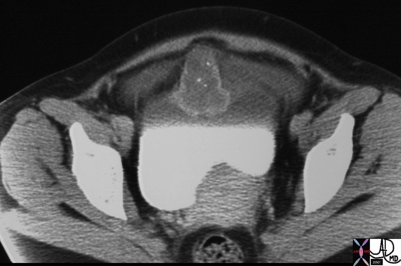
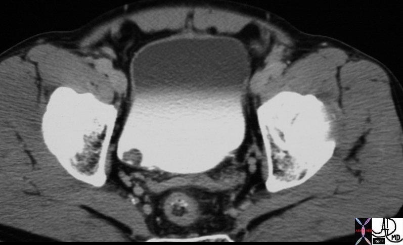
17102 |
| 17102 44F with hematuria fx anterior bladder mass fx calcification fx mass dx urachal adenocarcinoma imaging CTscan Courtesy Ashley Davidoff MD |
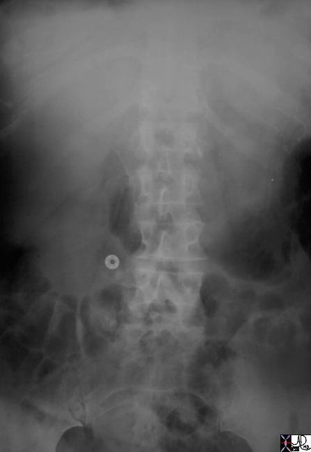
24636 |
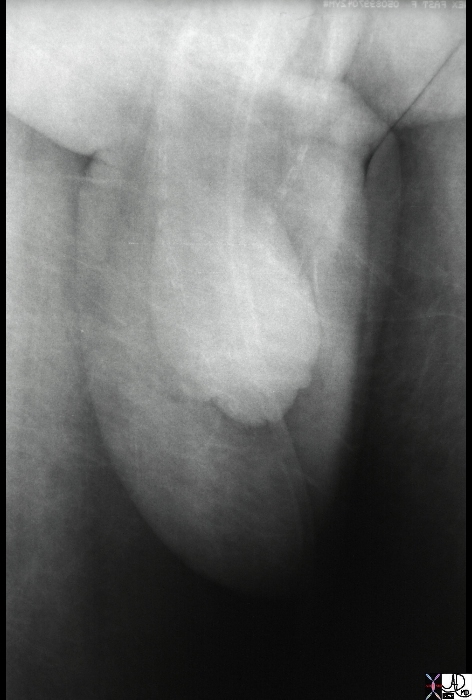
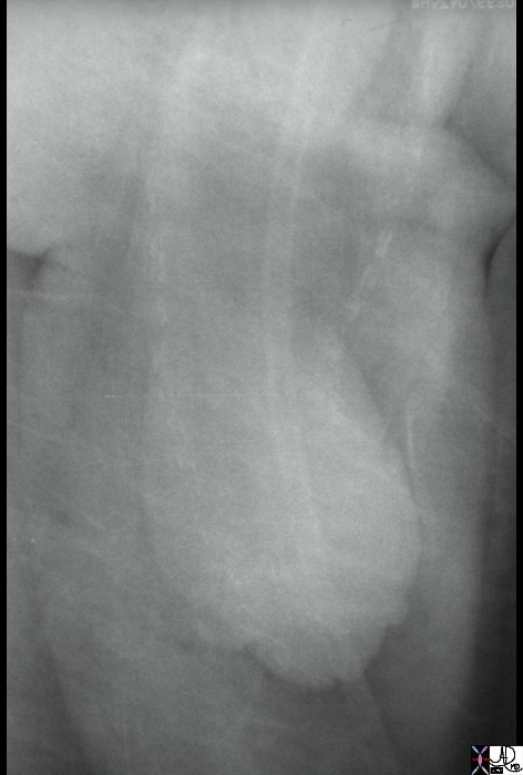
| 24636 penis vas deferens calcification diabetes age related sarcoidosis plain film X-ray Davidoff MD 24637 |
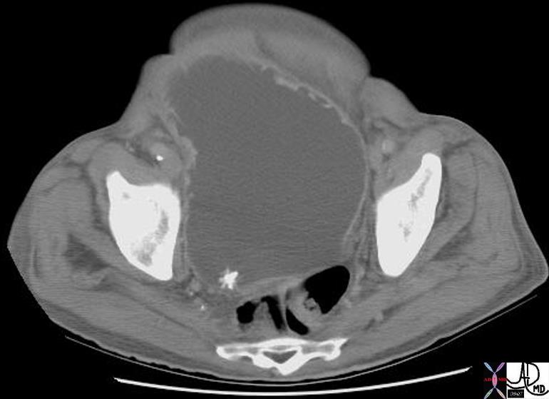
| 16568 |
| 16568 urinary bladder nodule fx relatively hypodense fx contrast in bladder dx bladder stone calculus CTscan Davidoff MD |
| 01313 |
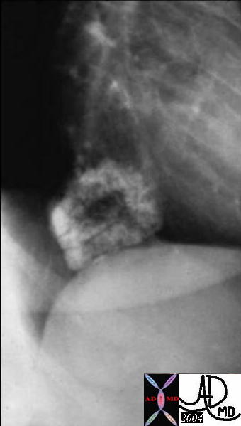
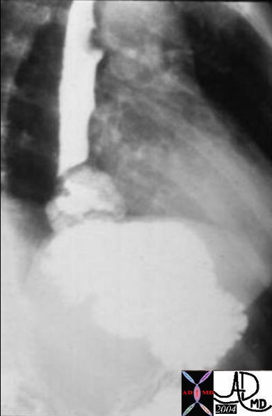
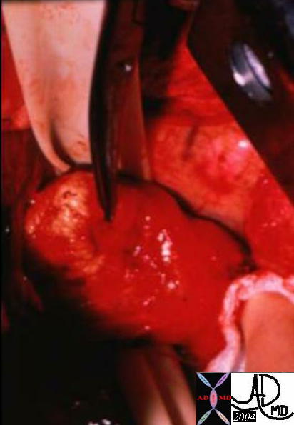
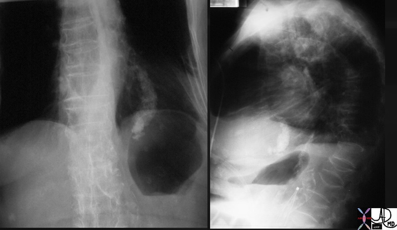
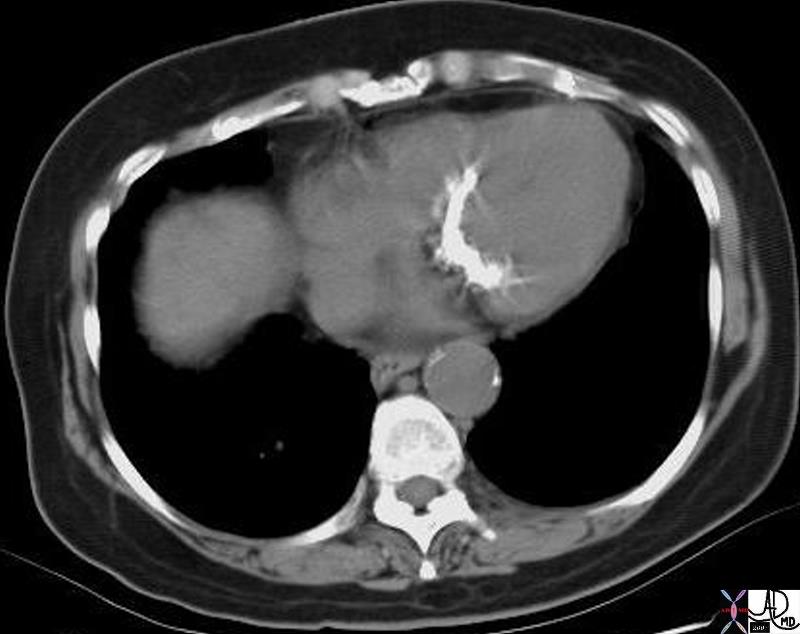
| Courtesy Barbara Banner MD 01313 code esophagus mass calcified calcification leiomyoma imaging radiology contrast X-Ray plain film CXR chest |
| 01312 |
| Courtesy Barbara Banner MD 01312 code esophagus mass calcified calcification leiomyoma barium swallow upper GI UGI imaging radiology contrast X-Ray |
| 01314 |
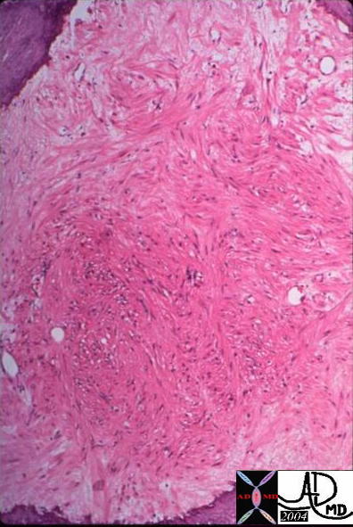
| Courtesy Barbara Banner MD 01314code esophagus mass calcified calcification leiomyoma grosspathology |
| 01316 |
| Courtesy Barbara Banner MD 01316 esophagus mass calcified calcification leiomyoma histopathology |
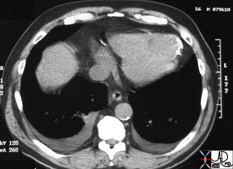
32744c01 |
| 32744c01 heart mitral valve mitral annulus mitral annular calcifcation MAC aortic caicifications osteopenia bone thoracic spine kyphosis compression fracture CXR plain film of the chest IVC filter Davidoff MD |
34130 |
| 34130 heart mitral valve mitral annulus mitral annular calcifcation MAC progression down the interventricular septum conduction defects nerve involvement CTscan Davidoff MD |
Calcification |
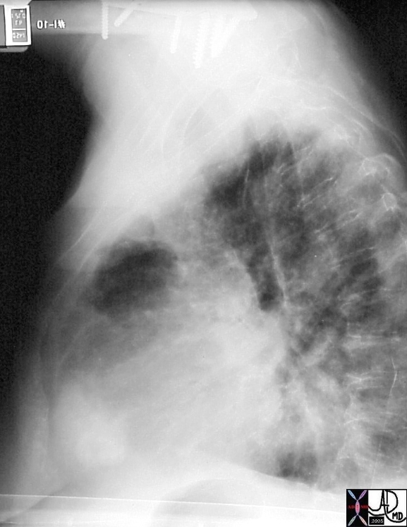
| 15537b01 hx 75F with left shoulder pain heart fx calcification ddx question calcific pericarditis probable dx metastatic chondrosarcoma imaging radiology plain film Courtesy Ashley Davidoff MD |
Calcification |
| 15537b03 hx 75F with left shoulder pain heart lung fx calcification fx parenchymal infiltrate fx pleural effusion dx small pulmonary infarct small Hamptons hump dx metastatic chondrosarcoma metastasis osteogenic sarcomatous malignant pulmonary emboli embolus imaging radiology CTscan Courtesy Ashley Davidoff MD |
15537b04d |
| 15537b04d hx 75F with left shoulder pain heart lung fx calcification fx parenchymal infiltrate fx pleural effusion dx small pulmonary infarct small Hamptons hump dx metastatic chondrosarcoma metastasis osteogenic sarcomatous malignant pulmonary emboli embolus imaging radiology CTscan Courtesy Ashley Davidoff MD |
MRI T2 weighted |
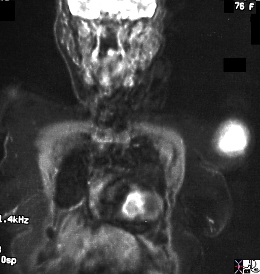
| 15551b01 bone heart cardiac pulmonary artery tumor pulmonary emboli 15537 T2 intensity over the heart T2 hyperintensity over left humerus shoulder MRIscan DxChondrosarcoma of the humerus metatstattic to the heart lungs Davidoff MD |
15537b07 |
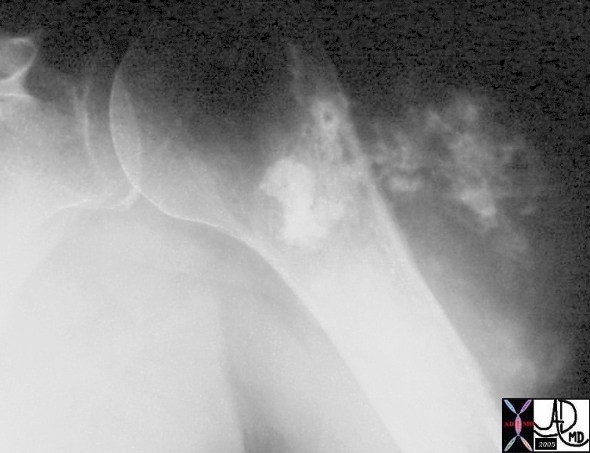
| 15537b07 hx 75F with left shoulder pain heart left humerus left shoulder fx cartilagenous popcorn type calcification dx chondrosarcoma chondrogenic sarcoma of the shoulder with metastasis to the RV right ventricle malignant imaging radiology plain film Courtesy Ashley Davidoff MD |
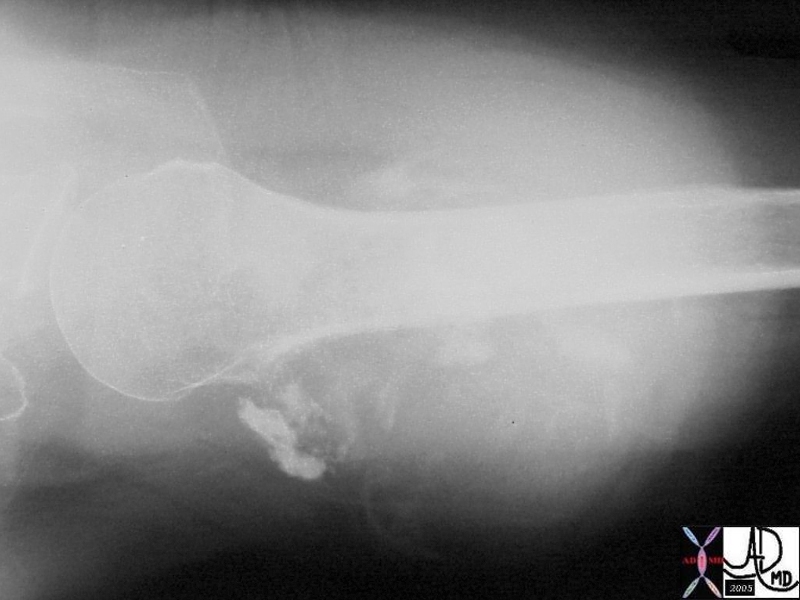
15537b08 |
| 15537b08 hx 75F with left shoulder pain heart left humerus left shoulder fx cartilagenous popcorn type calcification dx chondrosarcoma chondrogenic sarcoma of the shoulder with metastasis to the RV right ventricle malignant imaging radiology plain film Courtesy Ashley Davidoff MD |
Calcifications |
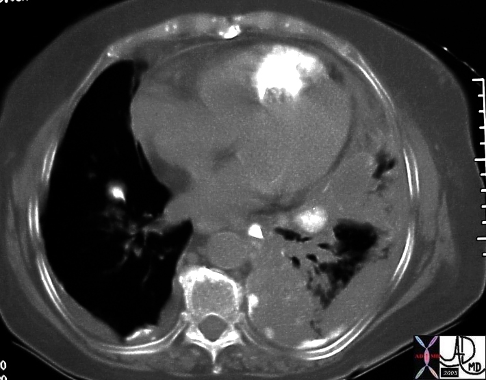
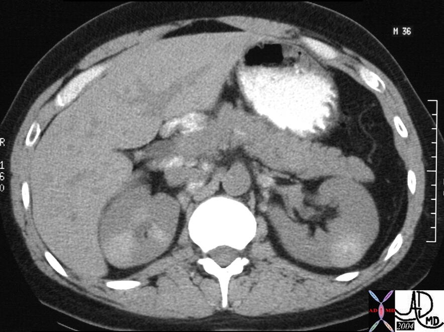
| 26477b02 Courtesy Ashley Davidoff MD pancreas anatomy lymph node lymphatic drainage fx calcified calcification dx sarcoidosis kidney mass calcification calcified imaging radiology CTscan |
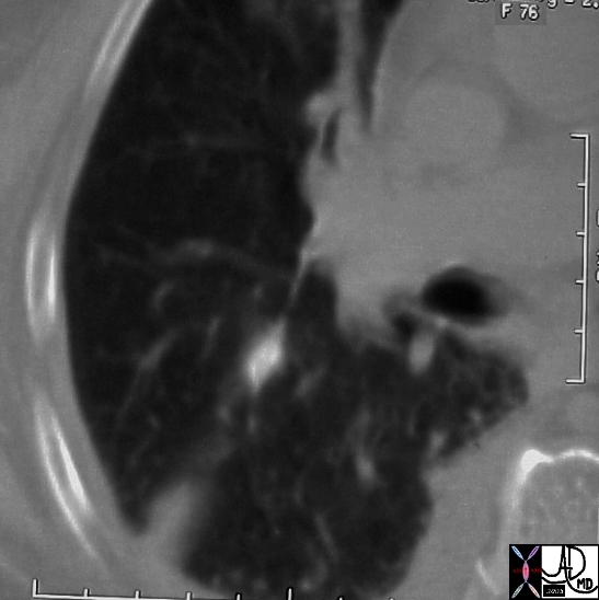
40406c |
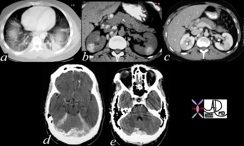
| 40406c Courtesy Ashley Davidoff MD code lung fx interstium fx thickening dx interstitial lung disease
ILD code pancreas abdomen lymph nodes fx calcified code brain meninges fx calcifications code dx sarcoidosis imaging radiology CT scan code inflammation immune granulomatous disease. Clinical involvement of the CNS is seen in approximately 5% of cases, though autopsy series report an incidence of up to 14%. It most commonly involves the basal meninges, cranial nerves, hypothalamus and pituitary gland. The CNS is the sole site of involvement in 1.5%. |
| 16568 |
| 16568 urinary bladder nodule fx relatively hypodense fx contrast in bladder dx bladder stone calculus CTscan Davidoff MD |
| 16758 |
| 16758 urinary bladder nodule fx relatively hypodense fx contrast in bladder dx bladder stone calculus CTscan Davidoff MD |
| 29600 |
| This single cross sectional view of the heart through the LV shows apical myocardial dystrophic calcification. The pericardium can be seen as a fine soft tissue density between the two layers of fat – the epicardial fat (inner layer) and the pericardial fat. This finding is diagnostic of a previous MI and may represent dystrophic calcification in the myocardium or within thrombus in an aneurysm of the apex. Courtesy Ashley Davidoff MD. 29600 code CVS cardiac heart MI apex calcification calcified IHD myocardium cardiac imaging radiology CTscan |

| 29601 |
| This single axial CT image shows a faint lucurvilinear lucency in the apical myocardium. This lucency is thought to be a fatty deposition in infarcted myocardial tissue. The calcification in the annulus is premature and unusual for this 56 year old male patient. Note the small bilateral pleural effusions. Courtesy Ashley Davidoff MD. 29601 code CVS cardiac heart MAC apical fat MI cardiac imaging radiology CTscan |
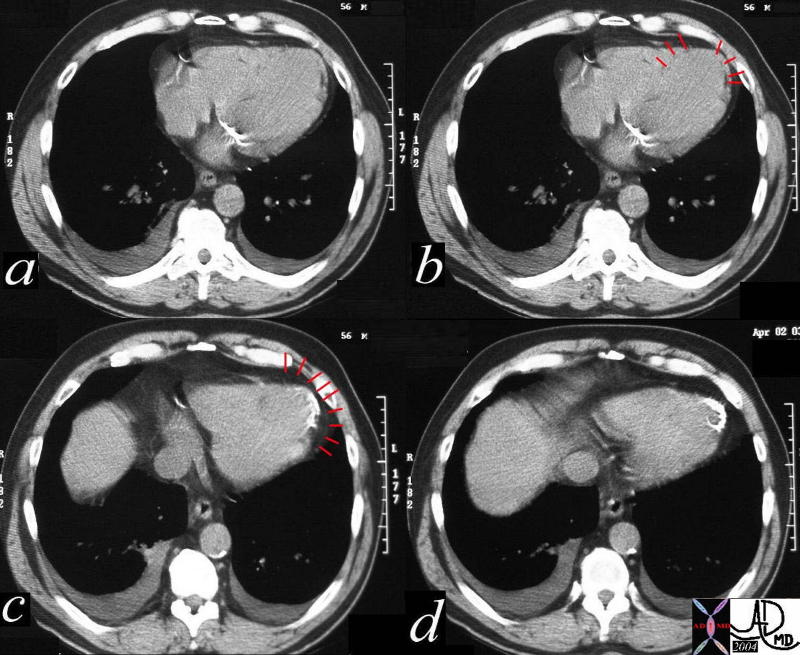
| 29601c01 |
| This series of CT image shows a faint curvilinear lucency in the apical myocardium (a,b) as well as myocardial calcification. The lucency identified by red lines in b, is thought to be a fatty deposition in infarcted myocardial tissue, and the calcification represents dystrophic calcification in the necrotic tissue. Note that the calcification is not in the pericardium which is identified by the red markers in c. The pericardium is suurounded by pericardial at on the outside and epicardial fat on the inside. Included in the differential diagnosis is an apical aneurysm with calcification in clot. The calcification in the annulus is premature and unusual for this 56 year old male patient. Note the small bilateral pleural effusions. Courtesy Ashley Davidoff MD. 29601c01 code CVS cardiac heart MAC apical fat MI calcium myocardium apex cardiac imaging radiology CTscan radiologists and detectives |
| 34305 |
| 34305 heart cardiac LV apex left ventricle fx calcification calcified coronary sinus fx enlarged DDX IHD rheumatic cardicitis embolism to LAD CTscan Davidoff MD 34305 34306 34298 34299 34300 |
| 34298 |
| 34298 heart cardiac MV mitral valve fx thickened LAE enlarged left atrium thickened chordae paillary muscle complex fx enlarged DDX IHD rheumatic cardicitis RHD Libman Sacks rheumatic heart disease CTscan Davidoff MD 34305 34306 34298 34299 34300 |
| 34299 |
| 34299 heart cardiac MV mitral valve fx thickened LAE enlarged left atrium thickened chordae paillary muscle complex fx enlarged DDX IHD rheumatic cardicitis RHD Libman Sacks rheumatic heart disease CTscan Davidoff MD 34305 34306 34298 34299 34300 |
| 22992 |
| 22992 liver + capsule + spleen small bowel stomach abdomen peritoneum peritoneal cavity fx mass + fx calcification + dx pseudomyxoma peritonei + imaging radiology CTscan Courtesy of Ashley Davidoff MD DB |
| 31118 |
| 31118 hx 75 F small bowel fx dilated fluid filled fx calcification dx SBO gallstone ileus CTscan Courtesy Ashley Davidoff MD DB |
| 31123 |
| 31123 hx 75 F small bowel fx dilated gallbladder fx calcifcation fx air dx SBO gallstone ileus CTscan Courtesy Ashley Davidoff MD DB |
| 26076 |
| 26076 Courtesy Ashley Davidoff MD code pancreas code bile duct + fx filling defects + fx calcifications + dx cholelithiasis + dx gallstone pancreatitis + imaging radiology CTscan C+ |
| 26079c01 |
| 26079c01 Courtesy Ashley Davidoff MD code pancreas + fx indurated + dx gallstone pancreatitis + imaging radiology CTscan C+ code bile duct + fx filling defects + fx calcifications + dx cholelithiasis + dx gallstone pancreatitis + imaging radiology |
| 20889 |
| 20889 code spleen + hilum + capsule + fx calcification + metastatic primary ovary + carcinoma + imaging radiology CTscan C- liver parenchyma capsule + fx calcification + nodule metastatic primary ovary ligamentum venosum fx calcium calcification dx metastatic primary ovary neoplasm malignant metastasis metastases cancer tumor skin third spacing pleural effusion |
| 16868 |
| 16868 diaphragm liver porta hepatis gastrohepatic ligament Glissons capsule + fx calcification + primary ovary + metastasis + imaging radiology CTscan neoplasm malignant carcinoma tumor cancer |
| 16869 |
| 16869 Punctate calcifications on the liver capsule pleura and diaphragm are noted on this chest CT. The patient has metastatic ovarian carcinoma to the pleura and pericardium with calcification. Courtesy Ashley Davidoff MD. code diaphragm liver porta hepatis gastrohepatic ligament Glissons capsule + fx calcification + primary ovary + metastasis + imaging radiology CTscan neoplasm malignant carcinoma tumor cancer spleen splenic |
| 32378.800 |
| 32378.800 urinary bladder calcifications stones wall forces gravity dependancy attached to the wall CTscan Davidoff MD 32378c01 |
| 32378c01 |
| 32378c01 urinary bladder calcifications stones wall forces supine prone gravity dependancy attached to the wall CTscan Davidoff MD 32378c01 32378.800 |
| 26853 |
| 26853 abdomen calcification linear IVC dx leiomyoma of the IVC KUB plain film of the abdomen Davidoff MD 26853 26858 26859 26854 26854b 26854.800 26851 26851b |
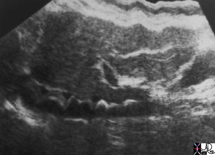
| 26859 |
| 26859 abdomen calcification linear wavy IVC USscan shadowing dx leiomyoma of the IVC Davidoff MD 26853 26858 26859 26854 26854b 26854.800 26851 26851b |
| 26854.800 |
| 26854.800 abdomen calcification linear IVC KUB plain film of the abdomen dx leiomyoma of the IVC Davidoff MD 26853 26858 26859 26854 26854b 26854.800 26851 26851b |
| 26851 |
| 26851 abdomen calcification linear IVC dx leiomyoma of the IVC inferior venacavagram venography Davidoff MD 26853 26858 26859 26854 26854b 26854.800 26851 26851b |
| 26864 |
| 26864 calcification linear IVC KUB plain film of the abdomen dx leiomyoma of the IVC path specimen X-Ray Davidoff MD 26853 26858 26859 26854 26854b 26854.800 26863 26851 26851b |