copyright 2008
Introduction
The principles of structure as outlined in the introduction persist into the principles in the evaluation of structure by imaging
These universal principles relate to the organelles of the cell, the cell itself, the tissues, organs and the body. The families, villages and towns are extensions of biological units and therefore are governed by similar principles. When studying the organelles and cells in cytology, it is useful to to try and use the same adjectives to describe the cell under the microscope, and structures identified in the operating room, on an X-ray or CTscan so that you appreciate that you are dealing with the universal biological unit- but just at a different level of complexity.
Parts
In the world that we can appreciate with our own eyes – from the organelles of the cells to the countries of our world, we can always break up structure into component parts. In the world of the atom and molecules, and in the world beyond our solar system, component parts are present as well. Parts are often subdivided in one of many ways but in the end there are the substantive components, and the connecting links that link the parts to each other and the parts to a bigger whole.
So for example the liver cells are held together by celleular bonds and connective tissue, and are linked to each other and the other organs by biliary radicles blood vessels nerves and lymphatics. These components at the microscopic level unite and form a liver lobule, which in turn unite in additive fashion to form lobes and finally the organ itself.
Size
The two sets of cells below represent some normal cells and malignant cells and one of the criteria to distinguish health against disease is the size of the cytoplasm and nucleus.
 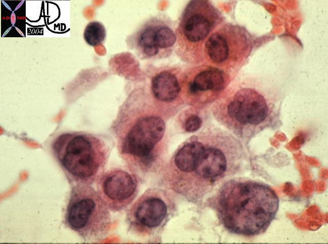
Which is the normal? The Nuclear-Cytoplasmic Ratio |
|
In this instance the relative size of the nucleus to the cytoplasm (nuclear cytoplasmic ratio = NC ratio) is markedly different. In the image on the left – the healthy cell – the overall size of the cytoplasm is relatively larger than the nucleus so that the NC ratio is normal whereas the nucleus is relatively large to the cytoplasm on the right image and hence the N-C ratio is large. The cells on the right are malignant liver cells and represent a primary hepatocellular carcinoma. Size of the cytoplasm and Size of the Nucleus (N-C ratio) 13440 liver hepatocytes cells cytology histology normal 5star Courtesy Barbara Banner MD 13447 liver HCC hepatocellular carcinoma cytopathology Courtesy Barbara Banner MD |
In the CXR below both the size and shape of the pulmonary artery are abnormal. Shape often is a reflection of a changing size of a structure. The bulging contour on the left heart border is the pulmonary artery and it is large becase of increased pressure in the pulmonary artery.
Shape
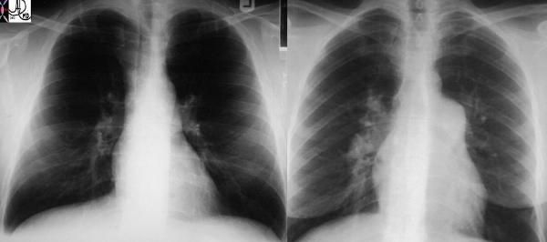
Normal and Abnormal Shape of the Heart |
| These two P-A chest X-rays show a normal cardiomediastinal image on the left and an enlarged main pulmonary artery (MPA) and right pulmonary artery (RPA) on the right in a patient with pulmonary hypertension. Courtesy Ashley Davidoff MD (22089 c) code lung main pulmonary artery MPA RPA large hypertension heart cardiac imaging plain film CXR |
As described shape can reflect a changing size because a sheer increase in volume or pressure in a structure will be reflected in the shape. The are many other factors that affect shape often caused by forces within the structure or the forces that surround the structure.
Position
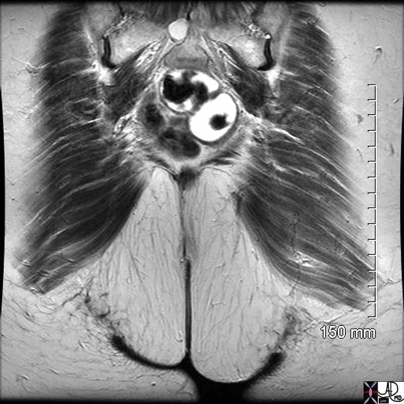
Position of structures in biology are often guided by an innate direction guided by the genes. For example below there is a normal patient on the left where the heart is in the left chest with its chambers normally positioned while the liver stomach and intestinal tract are positioned “normally” as well. In the image on the left a condition of situs inversus exists. The heart is on the right side, while the liver stomach and intestinal tract are positioned abnormally. Sometimes position is governed genetically, while other times it relates to the forces within the structure or surroundig the structure.
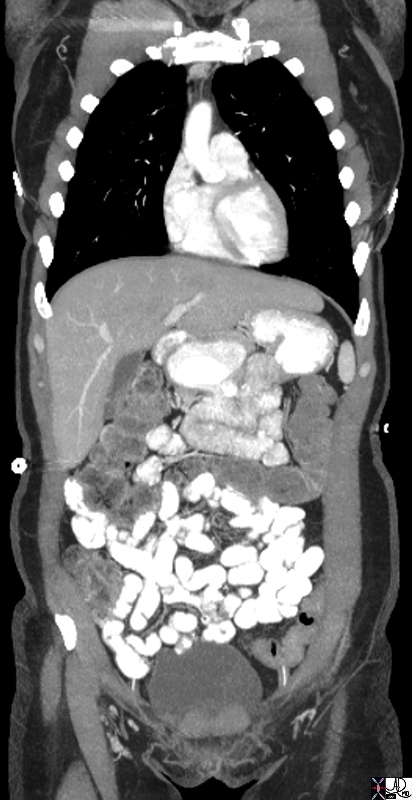 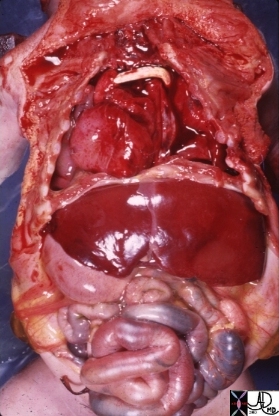
Normal and Situs Inversus |
| The Coronal image on the left shows normal position of the organs. The post mortem image is of a of a baby with situs inversus of the liver and the stomach associated with complex congenital heart disease s/p graft which can be seen in the anterior mediastinum. The nature of the heart disease is not known. Courtesy Ashley Davidoff MD. 10850 code cardiac heart situs inversus liver stomach heart bypass congenital grosspathology
60324 chest a abdomen pelvis heart gallbladder liver small bowel stomach situs solitus CTscan Coronal plane Davidoff MD |
The idea of space is key in this discussion on structural concepts. Each cell, tissue, and organ has to have space to work and to breathe. The position and relations of the units has to be optimal in order for them to be able to work and communicate with each other, and in the end come together as an empowered unit. The spatial organization of the liver described is a great example.
|
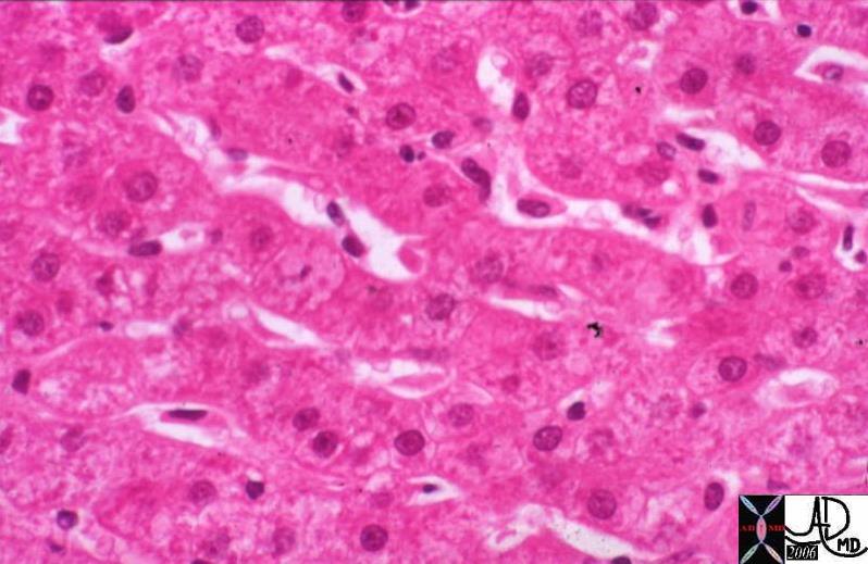
Positioning of Liver Cells in Cords |
|
The cells of the liver are organized in cords and plates and are organized like spokes of a wheel around the central vein. The plates and cords are lined by the sinusoids which are the channels which carry blood to the liver. Just below the sinusoids, between the wall of the sinusoid and the capsule of the liver there is a space called the space of Disse which carries the lymphatic fluid of the liver. (Image courtesy of Barbara Banner M.D.) 13445 |
The plates and cords are organized around the linear shape of the sinusoids and spaces of Disse, with the portal triads and central veins having their spatial arrangement to allow for optimal exposure of the liver cell surfaces to the circulation. We see this organizational pattern in our communities. There are industrial sites where the factoies are located and centered around access roads, ports or railway stations, and then we have our neighbourhoods where we live, centered around schools, playgrounds and grocery stores.
Our property lines are well defined and have to be respected. I had often wondered why cancer killed people and it came to me that cancer was a a disorder where property lines were not respected. Cancer cells are radicals and bullies in the community who have no respect for the community at large. They eat what they want to eat, provide no essential function, take all they can get, give nothing, disregard the rules, but most importantly they move into the space of others and take over, essentially throttling the well being of well meaning citizens of the community at large. The community cannot survive once their space and hence their function is taken over.
Character
Character is often the most difficult of the structural principles to grasp. In our inquisitive way we “taunt” structure by subjecting it to any one of the forces in our armamentarium and seeing how it reacts. We pull or push on it, throw different sorts of temperatures, chemical, radiational or magnetic fields toward it, and we observe its reaction. The reaction of the tissue to the force we subject it to is labelled its character. If one pulls the two ends of an elastic band apart and thereafter release them – they will return to their original position. It is thus characterized as having elastic properties. At the cellular level we stain the cells with different chemicals and observe how they react, at the clincial level we palpate and percuss, while at the imaging level we subject the tissues to ultrasound waves, X-rays, and changing magnetic forces in order to characterize them.
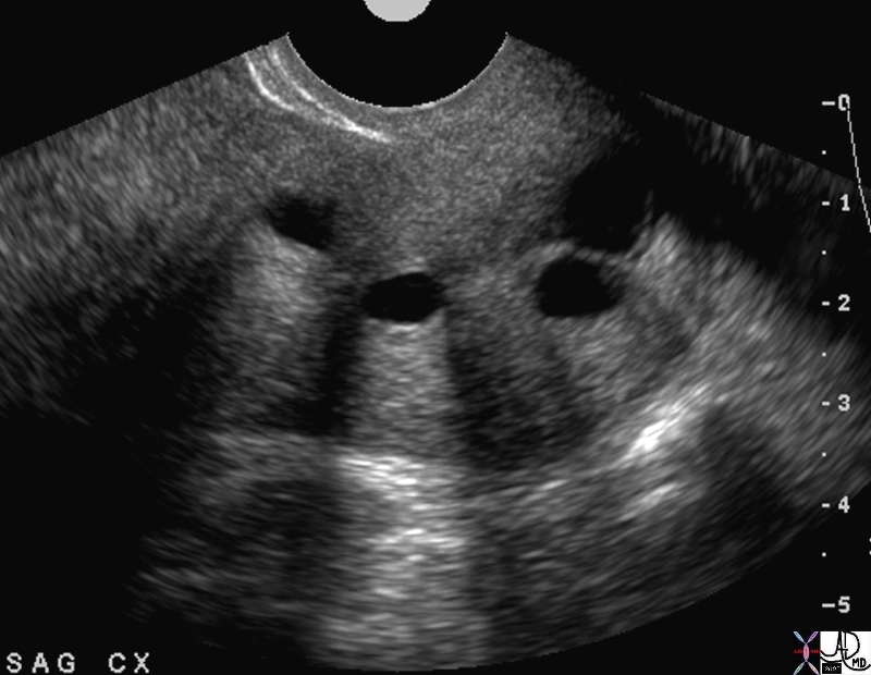
Classical Cysts by Ultrasound Anechoic, Backwall Enhancement and Through Transmission |
| The example of Nabothian cysts in the cervix characterised by ultrasound is noted above. Ultrasound waves are directed to the cervix via a transducer and they come into contact with the component tissues of the cervix. The waves are reflected and transmitted through solid tissue, so that the normal tissue has a homogeneous gray pattern, while the cysts present as black holes in the solid tissue. The sound waves are totally transmitted through the water of the cysts and without any reflection present a relatively powerful force to the back of the cyst. Back wall enhancement – a whiter back wall is best seen in cyst number two from the left. Behind the cysts the relative increase in power of the sound wave is seen as a wave of whiteness behind the cyst – “through transmission”. These three characterics, anechogenicity, back wall enhancement and through transmission characterise the structural abnormality as non concerning cysts.
49463 cervix fx cysts anechoic through transmission backwall enhancement dx Nabothian cysts USscan Davidoff MD |
Links Connections Relations
John Donne a clergyman and poet from the renaissance period stated that “… No man is an island …” In the same way no cell, organ, body or community is an island. Each is connected and dependant on the other members of the society in which they live. They are connected both structurally and functionally – an absolute necessity to survival.
The bonds take a variety of forms. The attachment of cells to each other is via terminal bars for example which are thoughto represent a polygonal bandat the periphery of each cell.. In the liver the cells have to align along a band of tissue in order to form the tubular sinusoids between them, and they are held togeher by a fine reticular network.
|
Positioning of Liver Cells in Cords – Reticulin Bond |
|
The cells of the liver are organized in cords and plates and are organized like spokes of a wheel around the central vein. The plates and cords are lined by the sinusoids which are the channels which carry blood to the liver. Just below the sinusoids, between the wall of the sinusoid and the capsule of the liver there is a space called the space of Disse which carries the lymphatic fluid of the liver. (Image courtesy of Barbara Banner M.D.) 13236 |

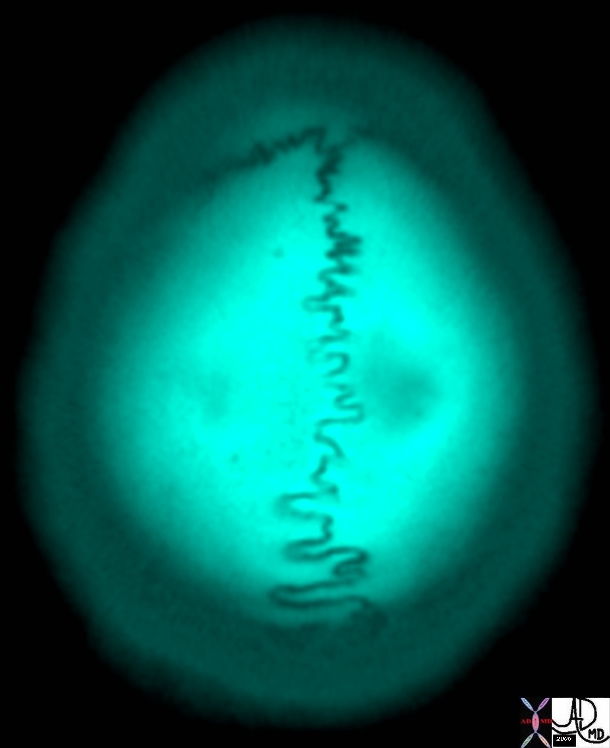
Sutures in the Skull |
| 46304b01 bone skull suture shape CTscan Davidoff art |
Small Vessels in the Skin and Muscle of the Buttock |
|
Note the linear and parallel orientation of the muscle fibres of the gluteus muscles (charcoal) and the reticular pattern of the connective tissue of the skin of the buttock (light gray) |

Cardiovascular System |
| 32368b05.800 cardiovascular system heart cardiac artery arteries capillary capillaries arterioles venules veins negative firces positive forces road highway TCV the common vein applied biology Davidoff art Davidoff MD |